It seems like everybody in the radiology field is now working with Artificial Intelligence (AI) in one way or another. Especially here in the Silicon Valley, the number of ventures that develop AI algorithms is increasing by the day. What are these newly developed algorithms focusing on? Most of them try to solve a specific problem, and they do that very well. There are many algorithms for detecting intracerebral haemorrhage on a CT scan of the brain. And maybe even more that detect lung nodules on a chest CT scan. These are all great advancements, but will they really change healthcare?
Yes, the aforementioned AI uses will provide benefit, but I think there is a whole different part of the radiology field that is even more important: improving the radiology workflow. This is where the big difference can be made. One essential aspect of workflow is the optimisation of image quality and reduction of radiation dose for CT images. So while AI can assist radiologists by diagnosing certain pathologies, it also has the potential to improve reconstruction of CT images. And not just improving image quality, but also reducing the radiation dose.
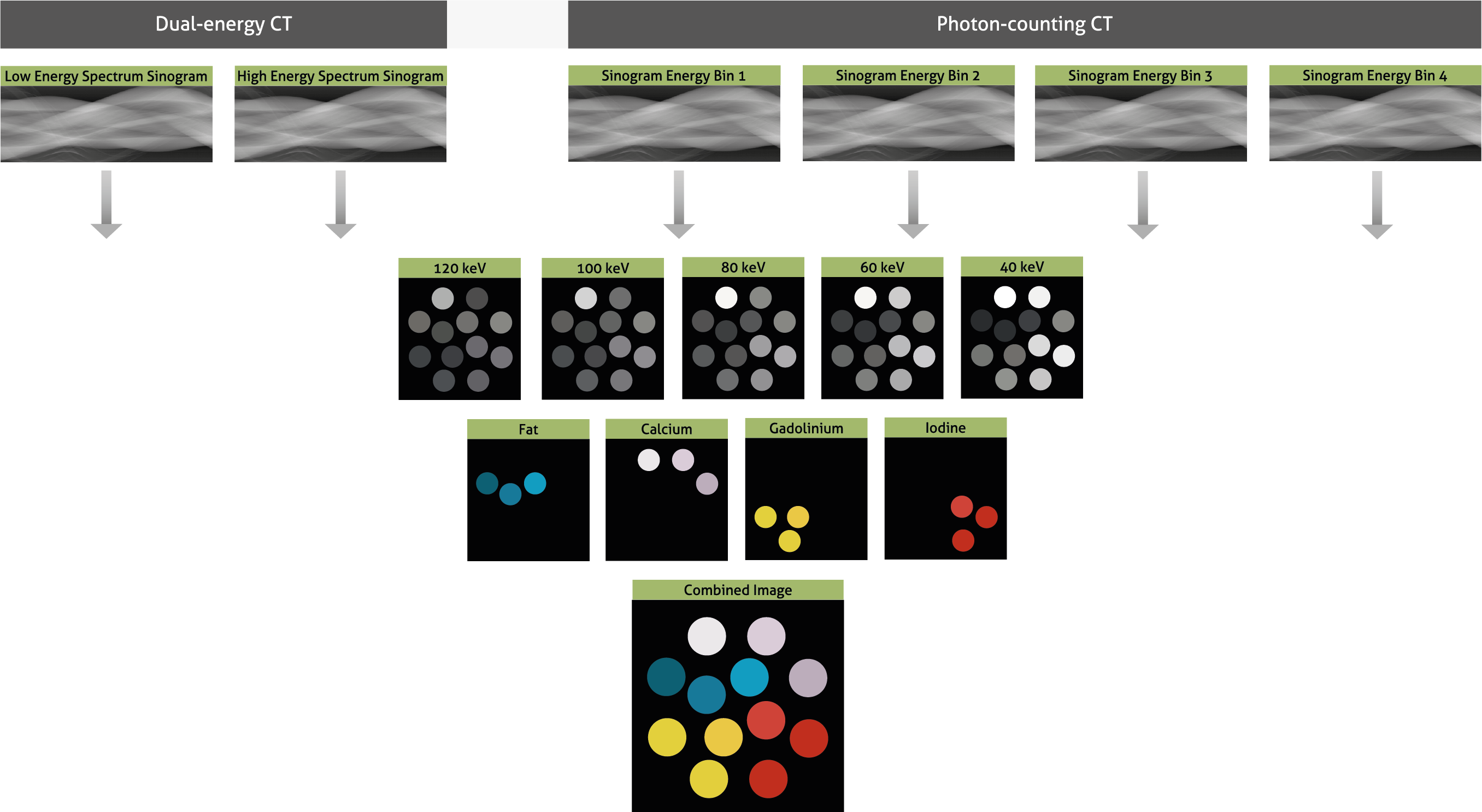
Recent studies have shown that routine-dose image quality can be created by training convolutional neural networks with low-dose CT images [1-3]. This may allow for a reduction of radiation dose [4-6] and reducing metal artifacts [7,8] while speeding up reconstruction-times. AI can also be used for optimisation of iterative reconstruction algorithms [9]. With upcoming technologies such as photon-counting CT, reconstruction of material-specific images is getting more and more complex. AI could allow for high-quality reconstructions of these complex data structures.
Read more about the evolution of reconstruction for CT images in the recently published article: “The evolution of image reconstruction for CT-from filtered back projection to artificial intelligence” [10].
Key Points:
- Advanced CT reconstruction methods are indispensable in the current clinical setting.
- IR is essential for photon-counting CT, phase-contrast CT, and dark-field CT.
- Artificial intelligence will potentially further increase the performance of reconstruction methods.
References
- Wolterink JM, Leiner T, Viergever MA, Isgum I (2017) Generative Adversarial Networks for Noise Reduction in Low-Dose CT. IEEE Trans Med Imaging 36:2536-2545
- Chen H, Zhang Y, Kalra MK et al (2017) Low-Dose CT With a Residual Encoder-Decoder Convolutional Neural Network. IEEE Trans Med Imaging 36:2524-2535
- Chen H, Zhang Y, Zhang W et al (2017) Low-dose CT via convolutional neural network. Biomed Opt Express 8:679-694
- Wu D, Kim K, El Fakhri G, Li Q (2017) Iterative Low-Dose CT Reconstruction With Priors Trained by Artificial Neural Network. IEEE Trans Med Imaging 36:2479-2486
- Chen Y, Liu J, Xie L et al (2017) Discriminative Prior – Prior Image Constrained Compressed Sensing Reconstruction for Low-Dose CT Imaging. Sci Rep 7:13868
- Kang E, Min J, Ye JC (2017) A deep convolutional neural network using directional wavelets for low-dose X-ray CT reconstruction. Med Phys 44:e360-e375
- Gjesteby L, Yang Q, Xi Y et al (2017) Reducing Metal Streak Artifacts in CT Images via Deep Learning: Pilot ResultsThe 14th International Meeting on Fully Three-Dimensional Image Reconstruction in Radiology and Nuclear Medicine, 1. Fully3D Community, Xi’an, pp 611-614
- Zhang Y, Yu H (2017) Convolutional Neural Network Based Metal Artifact Reduction in X-ray Computed Tomography. arXiv:170901581
- Kopp FK, Catalano M, Pfeiffer D, Rummeny EJ, Noël PB (2018) Evaluation of a machine learning based model observer for x-ray CT. Proc. SPIE, Medical Imaging 2018: Image Perception, Observer Performance, and Technology Assessment 10577:0S
- Willemink MJ, Noël PB (2018) The evolution of image reconstruction for CT-from filtered back projection to artificial intelligence. Eur Radiol. 2018 Oct 30.