Within the science community, the role of computed tomography (CT) for diagnosis is currently being debated. In this context, preliminary studies showed that chest CT imaging of the lung provides improved sensitivity when associated with RT-PCR for individuals suspected of having COVID-19 [1]. The primary features seen on a lung affected by COVID-19 are peripheral focal or multi-focal ground-glass opacities, consolidations and crazy-paving patterns.
Non-contrast chest CT has been useful not only to detect, quantify the severity and assess the progression of the disease, but also to evaluate the potential response to therapy alternatives.
In the current situation of improving COVID-19 therapy, radiologists face the challenge that more and more cases must be read, annotated and prioritized. An AI-powered analysis of radiological images has the potential to reduce this growing burden on radiologists and speed up their reading time.
Collaboration as an answer to the complex situation
To support the fight against the COVID-19 pandemic, Siemens Healthineers worked closely together with collaboration partners and inhouse expert teams to jointly focus on the development of a new algorithm [2]. With the AI research and development team in Princeton, NJ, USA, the software development center in Bangalore, India, CT product experts in Forchheim, Germany, customer collaboration partners in Paris, France and the power of the Sherlock supercomputer, Siemens Healthineers is enhancing their AI portfolio with an algorithm dedicated to CT imaging.
Learn more about the collaboration by clicking here.
CT Pneumonia Analysis algorithm [2] – how does it work?
The algorithm is designed to automatically identify and quantify abnormal tomographic patterns in the lungs from chest CT for research purposes.
The system takes a non-contrasted chest CT as input, identifies and 3D segments the lungs and lobes before segmenting the abnormalities. It quantifies both the extent of COVID-19 abnormalities and presence of high opacities. High opacity abnormalities were shown to correlate with severe symptoms.
The computed results could be used to analyze the severity and the progression of abnormalities in patients exhibiting COVID-19 symptoms.
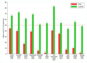
The performance of the method is evaluated on a database of 100 COVID-19 cases and 100 controls from multiple institutions from Canada, Europe and the U.S. Ground truth is established by computing the same measures from manual annotations of the lesions, lungs and lobes.
Learn more about how the CT Pneumonia Analysis [2] algorithm works and how you can use it for free!
Disclaimer:
[1] https://pubs.rsna.org/doi/10.1148/radiol.2020201365, 22.04.2020
[2] For Research Use Only. Not for use in diagnostic procedures.