Continuing our series from last week, where we get an inside look at the topic of artificial intelligence (AI) in relation to radiological journals over three brief interviews with the Editors-in-Chief of the European Society of Radiology’s three flagship journals, we speak with the Editor-in-Chief of Insights into Imaging, Professor Luis Martí-Bonmatí. Professor Martí-Bonmatí is the director of the Medical Imaging Department at La Fe University and Polytechnic University Hospital, as well as the Chief of Radiology at Quirónsalud Hospital, and resides in Valencia, Spain.
What Artificial Intelligence-related trends are you seeing in the field of radiology at the moment?
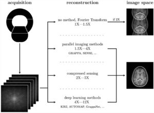
From my perspective, there are three main aspects where AI is impacting the way we practice radiology: image acquisition, organ segmentation, and tissue properties extraction. Although AI will be used to improve the rest of the workflow, such as with imaging appropriateness criteria, patient scheduling, and protocol definition, radiation dose reduction and image quality improvements, image reconstruction from minimal raw data (as a partial “virtual scan”), organ segmentation (which can be defined as “virtual dissection”), and tissue properties extraction (which also can be recognized as “virtual biopsy”) are clearly recognized as quite relevant tools to improve the quality of the radiological report and to ease the work of radiologists while reducing patient scanning times. In this same way, AI-derived lesion detection and characterization are “partial” aspects of the virtual dissection and biopsy approaches.
Which topics and (clinical) questions within Artificial Intelligence are currently the most prominent in your journal (e.g. Machine Learning, Convolutional Neural Networks, computer-aided diagnosis, etc.)?
Up to now, most papers on AI published in Insights Into Imaging are position papers and general education reviews on how machine learning, deep learning, and convolutional neural networks can help technicians and radiologists. We are also in the process of publishing papers on the ethics and impact on the clinical decision-making process of AI solutions. There is a growing trend to discuss which are the optimum metrics to validate both imaging biomarkers and AI algorithms. Aspects like the appropriateness of area under the curve (AUC) to measure AI accuracy and finding new metrics closer to clinical impact and application are a matter of relevancy. Of course, we appreciate any paper related to critical reviews on how radiologists should be involved in these unique AI opportunities to improve imaging workflow and analyze patient images objectively.
In which area do you think AI is quite advanced, and in which areas will further research be needed?
Value-based radiology is governing what radiologists perform these days. AI will influence all aspects of our value-chain. In my view, image appropriateness and image utilization, scheduling time and location, image protocol and safety issues, image acquisition, radiation dose, and image quality evaluations will surely benefit from AI. Structured reporting and radiomic features extraction from segmented organs and lesions will also help radiologists to integrate all the available information for the benefit of the patient (grading, staging, predicting, and prognostication). Deep learning and neural networks will allow radiologists’ time and effort to be more efficient, providing time to be involved in clinical duties such as multidisciplinary discussions, image guiding treatments, clinical and experimental research, and clinical innovation steps. Finally, less-stressed radiologists will be more deeply involved in imaging-related improvements in the healthcare cycle, fighting against professional burnout.


