Throughout this series, we will get an inside look at the topic of artificial intelligence (AI) in relation to radiological journals over three brief interviews with the Editors-in-Chief of the European Society of Radiology’s three flagship journals: European Radiology (Professor Yves Menu), European Radiology Experimental (Professor Francesco Sardanelli), and Insights into Imaging (Professor Luis Martí-Bonmatí). This week, we begin with Professor Yves Menu, Editor-in-Chief of European Radiology, who has been Editor-in-Chief since 2017 and currently resides in Paris, France.
What Artificial Intelligence-related trends are you seeing in the field of radiology at the moment?
Globally, we see an increasing proportion of submissions dealing with artificial intelligence (AI). Today, they account for 20-30% of all submissions. I expect that we will reach 50% in the near future; however, AI will not necessarily be the main topic of the study, but conversely used as a method of image analysis.
Which topics and (clinical) questions within artificial intelligence are currently the most prominent in your journal (e.g. machine learning, convolutional neural networks, computer-aided diagnosis, etc.)?
 Machine learning, convolutional neural networks, etc. are tools, not clinical questions; they are tentative methods to answer these questions. I would say that today, we are half-way between technical considerations, such as “which is the best machine learning algorithm for image analysis”, and clinical questions, such as “how can this method help me predict a tumor grade or lymph node involvement”. Most manuscripts explore the possibility of AI and try to validate specific methods to achieve a clinical task; however, these papers are still very focused. Researchers are working hard to improve their methodology; for example, enlarging samples as much as possible, clarifying algorithms, and producing relevant validation. This rather technical step will remain necessary until our readership becomes familiar with the new vocabulary and learns how to evaluate the robustness of publications by themselves.
By the way, this again stresses the importance of multicentric cooperation for validation, as the machines faithfully reproduce the human work in the sense that each center defines its own “subjective truth”, not necessarily identical to that of another institution.
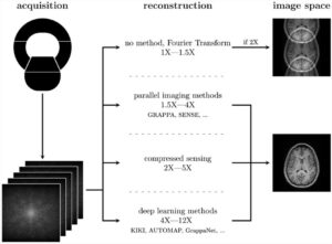
The vast majority of submissions deal with image analysis using non-visible data embedded within images, and/or comparison with a large database, in order to analyze the image better than a human could. An emerging question is whether or not AI needs images to analyze the signal. Images are reconstructed for the human eye, not for the machine. What if the machine can analyze raw data? However, there are other applications, starting with the improvement of image production, at the reconstruction level, opening a new perspective in further reducing the radiation dose while improving the image quality for the human eye.
Finally, we start seeing some attempts to use AI in order to improve our organization. If it is still at the very beginning in our environment, it is already a prominent issue outside the medical world.
In which area do you think AI is quite advanced, and in which areas will further research be needed?
I don’t know about any field that would not need further research. AI obviously needs further research – and in all directions.
From a methodological point of view, AI methods should be clarified and shared. Regarding statistics, and even if all of us are not, by far, expert statisticians, we have some common basic knowledge, like significance level or intraclass correlation, for example. AI methods still need to be better understood and shared, and probably standardized. We are not yet there.
Clinical applications are promising; however, they are still very focused. AI methods need to widen their scope and probably be integrated with basic tools. Do we need a program that only predicts lymph nodes’ involvement of squamous cell head and neck cancers? Probably not, as we would ask the AI to help us on a more global scale for the diagnosis and the management of these tumors, including detection, characterization, prognosis evaluation, and response to treatment.
In the near future, we can expect the integration of various solutions into a limited number of software programmes. Day after day, we are approaching the conclusion that AI has the potential to become the most efficient assistant we could ever dream of. Conversely, we will have to learn how to remain the ‘Master of Ceremony’.


