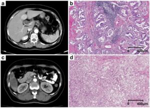
This pilot study aims to investigate whether liver fibrosis can be staged by deep learning techniques based on CT images. It included CT examinations of patients who underwent dynamic contrast-enhanced CT for evaluations of the liver and for whom histopathological information regarding liver fibrosis stage was available. Some additional images for training data were generated by rotating or parallel shifting the images or adding Gaussian noise and supervised training was used to minimise the difference between the liver fibrosis stage and the fibrosis score obtained from deep learning based on CT images (FDLCT score) output by the model. The results concluded that liver fibrosis can be staged by using a deep learning model based on CT images, with moderate performance.
Key Points:
- Liver fibrosis can be staged by a deep learning model based on magnified CT images including the liver surface, with moderate performance.
- Scores from a trained deep learning model showed moderate correlation with histopathological liver fibrosis staging.
- Further improvement are necessary before utilisation in clinical settings.
Article: Deep learning for staging liver fibrosis on CT: a pilot study
Authors: Koichiro Yasaka, Hiroyuki Akai, Akira Kunimatsu, Osamu Abe and Shigeru Kiryu


