








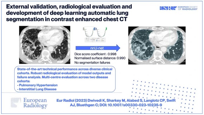
The authors of this study developed a 3D nnU-Net-based model for automatic lung segmentation in computed tomography pulmonary angiography (CTPA) imaging that was found to be highly accurate, clinically evaluated, and externally tested in patient cohorts with a spread of lung disease.
Key points
- Accurate, externally validated CT pulmonary angiography (CTPA) lung segmentation model tested in two large heterogeneous clinical cohorts (pulmonary hypertension and interstitial lung disease).
- No segmentation failures and robust review of model outputs by radiologists found 1 (0.5%) clinically significant segmentation error.
- Intended clinical use of this model is a necessary step in techniques such as lung volume, parenchymal disease quantification, or pulmonary vessel analysis.
Authors: Krit Dwivedi, Michael Sharkey, Samer Alabed, Curtis P. Langlotz, Andy J. Swift & Christian Bluethgen