Impact of signal intensity normalization of MRI on the generalizability of radiomic-based prediction of molecular glioma subtypes

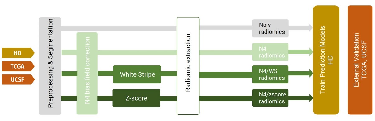
With the help of radiomics, standard medical images can be transformed into detailed, high-dimensional data sets that go beyond what the eye can see. The typical workflow of radiomic projects involves a series of sequential processes, including image registration, intensity normalization, and segmentation of the region of interest. While there is a general agreement on the essential steps, consensus on the best preprocessing methods remains elusive. Numerous works have highlighted the promising role of radiomics in the field of neuro-oncology, such as in predicting patient survival, evaluating treatment responses, and pinpointing key biomarkers.

In our article “Impact of signal intensity normalization of MRI on the generalizability of radiomic-based prediction of molecular glioma subtypes”, published in European Radiology, we delve into the pivotal role of MRI signal intensity normalization in boosting the accuracy and generalizability of radiomic-based models for identifying molecular subtypes of gliomas. Intensity normalization standardizes the range and scale of image pixel intensities, reducing variability between scans.

Our study reveals that intensity normalization approaches like WhiteStripe and Z-score normalization are foundational elements that significantly contribute to the robustness and generalizability of radiomic-based machine learning models. Specifically, while intensity normalization was not relevant when applying the developed radiomic-based machine-learning model to homogeneous data from the same institution, it was crucial to preserve the model performance when used in an external heterogeneous, multi-institutional setting.

With our work, we uncover the importance of MRI signal intensity normalization to address the generalization gap of radiomic-based machine learning models and thereby facilitate its clinical translation.

Key points

  • MRI-intensity normalization increases the stability of radiomics-based models and leads to better generalizability.
  • Intensity normalization did not appear relevant when the developed model was applied to homogeneous data from the same institution.
  • Radiomic-based machine learning algorithms are a promising approach for simultaneous classification of IDH and 1p/19q status of glioma.

Article: Impact of signal intensity normalization of MRI on the generalizability of radiomic-based prediction of molecular glioma subtypes

Authors: Martha Foltyn-Dumitru, Marianne Schell, Aditya Rastogi, Felix Sahm, Tobias Kessler, Wolfgang Wick, Martin Bendszus, Gianluca Brugnara & Philipp Vollmuth

WRITTEN BY

  • Martha Foltyn-Dumitru

    Department of Neuroradiology, Heidelberg University Hospital, Heidelberg, Germany; Section for Computational Neuroimaging, Department of Neuroradiology, Heidelberg University Hospital, Heidelberg, Germany

  • Philipp Vollmuth

    Department of Neuroradiology, Heidelberg University Hospital, Heidelberg, Germany; Section for Computational Neuroimaging, Department of Neuroradiology, Heidelberg University Hospital, Heidelberg, Germany; Division of Medical Image Computing (MIC), German Cancer Research Center (DFKZ), Heidelberg, Germany

Latest posts

Become A Member Today!

You will have access to a wide range of benefits that can help you advance your career and stay up-to-date with the latest developments in the field of radiology. These benefits include access to educational resources, networking opportunities with other professionals in the field, opportunities to participate in research projects and clinical trials, and access to the latest technologies and techniques. 

Check out our different membership options.

If you don’t find a fitting membership send us an email here.

Membership

for radiologists, radiology residents, professionals of allied sciences (including radiographers/radiological technologists, nuclear medicine physicians, medical physicists, and data scientists) & professionals of allied sciences in training residing within the boundaries of Europe

  • Reduced registration fees for ECR 1
  • Reduced fees for the European School of Radiology (ESOR) 2
  • Exclusive option to participate in the European Diploma. 3
  • Free electronic access to the journal European Radiology 4
  • Content e-mails for all ESR journals
  • Updates on offers & events through our newsletters
  • Exclusive access to the ESR feed in Juisci

€ 11 /year

Yes! That is less than €1 per month.

Free membership

for radiologists, radiology residents or professionals of allied sciences engaged in practice, teaching or research residing outside Europe as well as individual qualified professionals with an interest in radiology and medical imaging who do not fulfil individual or all requirements for any other ESR membership category & former full members who have retired from all clinical practice
  • Reduced registration fees for ECR 1
  • Free electronic access to the journal European Radiology
  • Content e-mails for all 3 ESR journals 4
  • Updates on offers & events through our newsletters
  • Exclusive access to the ESR feed in Juisci

€ 0

The best things in life are free.

ESR Friends

For students, company representatives or hospital managers etc.

  • Content e-mails for all 3 ESR journals 4
  • Updates on offers & events through our newsletters

€ 0

Friendship doesn’t cost a thing.

The membership type best fitting for you will be selected automatically during the application process.

Footnotes:

01

Reduced registration fees for ECR 2024:
Provided that ESR 2023 membership is activated and approved by August 31, 2023.

Reduced registration fees for ECR 2025:
Provided that ESR 2024 membership is activated and approved by August 31, 2024.

02
Not all activities included
03
Examination based on the ESR European Training Curriculum (radiologists or radiology residents).
04
European Radiology, Insights into Imaging, European Radiology Experimental.