Dr. Rizwan Aslam, of the University of California, San Francisco (UCSF), presented an abstract at RSNA in 2008 which showed that it was possible to screen for osteoporosis from CT colonoscopy scans while I was a resident at UCSF. Subsequently, other papers showed that there were strong correlations between CT measurements and the measurements obtained from dual-energy x-ray absorptiometry (DXA). This was an important finding because the development of low bone mineral density (BMD) is a pernicious process and is sometimes only detected after the development of fractures. In addition, most patients who are at risk for osteoporosis and osteopenia aren’t always screened using DXA. Recent papers showed that the CT attenuation of L1 was a reasonable choice for opportunistic screening because it is often unaffected by degenerative changes of the spine and had good performance in the detection of osteoporosis and osteopenia. However, T12, L1, and L2 are some of the most fractured spinal vertebrae, which limits using the CT attenuation of L1 in a few patients.
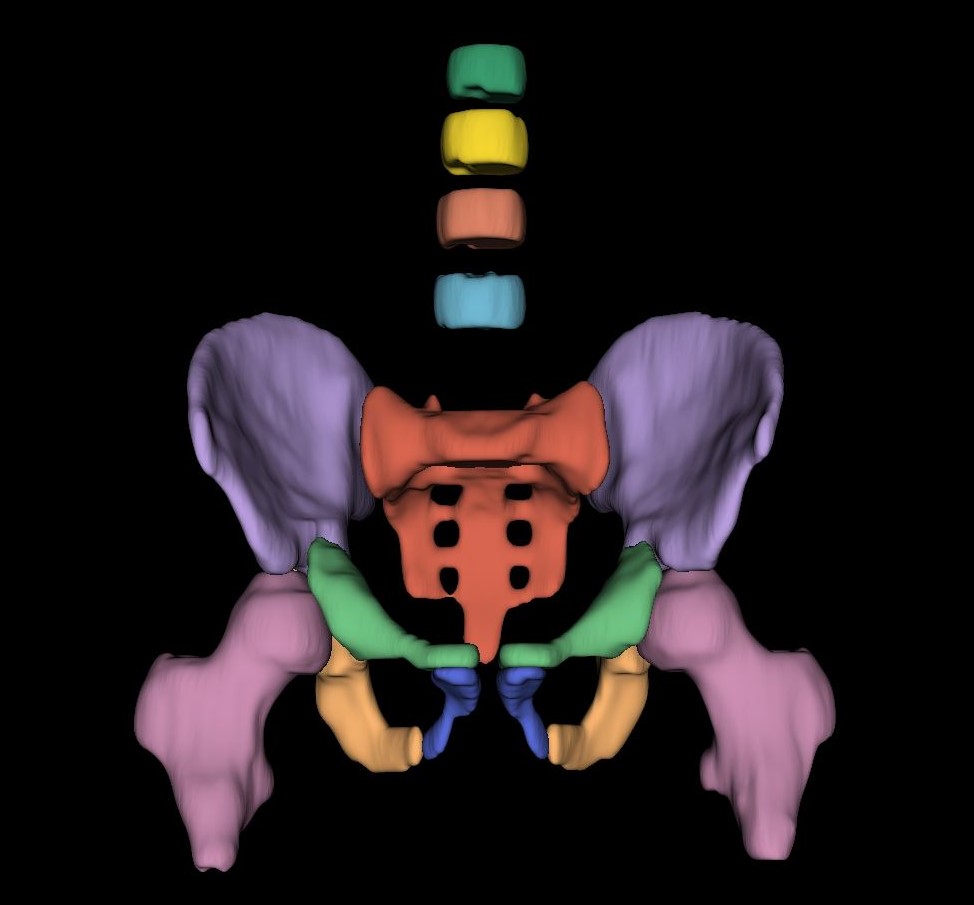
In a CT scan of the abdomen and pelvis, several bones are available for analysis. We hypothesized that the CT attenuation of all bones available would be more informative than the CT attenuation of a single bone (L1). This is analogous to reading an entire book versus a single page – reading the entire book, of course, provides a better understanding.
We found that using the data from all bones available on CT scans of the abdomen and pelvis was more informative.
Key points
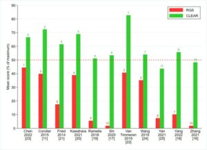
- Multivariable SVM model using the CT attenuation of multiple bones and clinical/demographic data was more predictive than using the CT attenuation at L1 only.
Authors: Ronnie Sebro & Cynthia De la Garza-Ramos