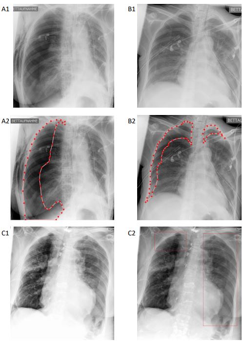
Due to the noisy annotation quality of public training data and confounding thoracic tubes, the diagnostic accuracy of artificial intelligence (AI) pneumothorax (PTX) detection in chest radiographs is limited. Therefore, the authors of this study hypothesized that in-image annotations of the dehiscent visceral pleura for algorithm training boosts the algorithm’s performance and suppresses confounders. Their results in this study are aimed at drawing attention to the necessity of high-quality in-image localization in training data in order to reduce the risks of unintentionally biasing the training process of pathology-detecting AI algorithms.
Key points
- Established pneumothorax-detecting artificial intelligence algorithms trained on public training data are strongly limited and biased by confounding thoracic tubes.
- We used high-quality in-image annotated training data to effectively boost algorithm performance and suppress the impact of confounding thoracic tubes.
- Based on our results, we hypothesize that even hidden confounders might be effectively addressed by in-image annotations of pathology-related image features.
Authors: Johannes Rueckel, Christian Huemmer, Andreas Fieselmann, Florin-Cristian Ghesu, Awais Mansoor, Balthasar Schachtner, Philipp Wesp, Lena Trappmann, Basel Munawwar, Jens Ricke, Michael Ingrisch & Bastian O. Sabel