Radiomics based on brain MRI is expected to be a useful tool for early identification and prediction of WMH progression

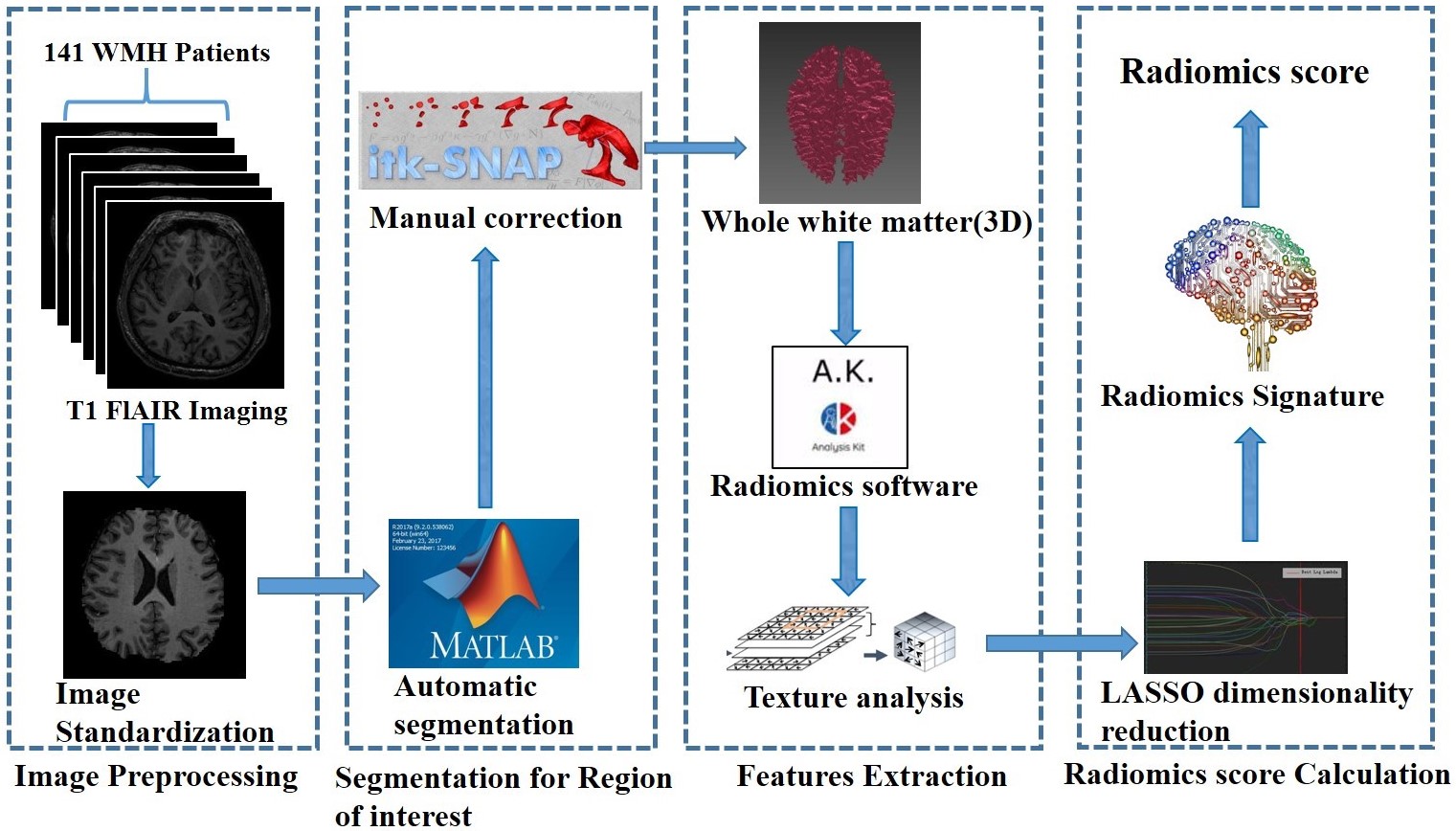
As a research hotspot in recent years, radiomics provides a new perspective for image diagnosis or evaluation by mining a large number of non-traditional visual information in medical images and adds new indicators (image markers). Radiomics may become a non-invasive, low-cost and easy to popularize imaging tool. White matter hyperintensity (WMH) refers to the high signal of white matter area on T2WI or flair MRI images. It is a common finding of brain MRI in the elderly and it is an important part of cerebral small vessel disease (cSVD). WMH is a growing pathological change in an ageing society. It is considered to be a risk factor for cognitive decline, dementia, depression, stroke, gait and balance problems.

In the present study, we used radiomics to predict the progress of WMH and the related risk factors, hoping to find a way for early detection and early diagnosis of WMH, and provide a time window for early intervention, containment, and slowing down the progress of white matter. Based on our study, we conclude that radiomics can well predict the progression of white matter lesions and age, blood lipid, and BMI are the main factors for the progression of white matter lesions. In conclusion, radiomics based on brain MRI is expected to be a useful tool for early identification and prediction of WMH progression. Early intervention strategies such as controlling blood lipid and lowering BMI may reduce the occurrence, and delay the progression, of WMH.

Key points

  • Radiomics features detected by magnetic resonance imaging may be used to predict the progression of white matter hyperintensities.
  • Radiomics may be used to identify risk factors associated with the progression of white matter hyperintensities.
  • Radiomics may serve as non-invasive biomarkers to monitor white matter status.

Article: Radiomics from magnetic resonance imaging may be used to predict the progression of white matter hyperintensities and identify associated risk factors

Authors: Zhenyu Shu, Yuyun Xu, Yuan Shao, Peipei Pang & Xiangyang Gong

WRITTEN BY

  • Yuyun Xu

    Department of Radiology, Zhejiang Provincial People’s Hospital; The Affiliated People’s Hospital of Hangzhou Medical College, Hangzhou, China

  • Xiangyang Gong

    Department of Radiology, Zhejiang Provincial People’s Hospital; The Affiliated People’s Hospital of Hangzhou Medical College, Hangzhou, China

Latest posts

Become A Member Today!

You will have access to a wide range of benefits that can help you advance your career and stay up-to-date with the latest developments in the field of radiology. These benefits include access to educational resources, networking opportunities with other professionals in the field, opportunities to participate in research projects and clinical trials, and access to the latest technologies and techniques. 

Check out our different membership options.

If you don’t find a fitting membership send us an email here.

Membership

for radiologists, radiology residents, professionals of allied sciences (including radiographers/radiological technologists, nuclear medicine physicians, medical physicists, and data scientists) & professionals of allied sciences in training residing within the boundaries of Europe

  • Reduced registration fees for ECR 1
  • Reduced fees for the European School of Radiology (ESOR) 2
  • Option to participate in the European Diploma. 3
  • Free electronic access to the journal European Radiology 
  • Content e-mails for all ESR journals4
  • Updates on offers & events through our newsletters
  • Exclusive access to the ESR feed in Juisci

€ 11 /year

Yes! That is less than €1 per month.

Free membership

for radiologists, radiology residents or professionals of allied sciences engaged in practice, teaching or research residing outside Europe as well as individual qualified professionals with an interest in radiology and medical imaging who do not fulfil individual or all requirements for any other ESR membership category & former full members who have retired from all clinical practice
  • Reduced registration fees for ECR 1
  • Option to participate in the European Diploma. 3
  • Free electronic access to the journal European Radiology
  • Content e-mails for all ESR journals 4
  • Updates on offers & events through our newsletters
  • Exclusive access to the ESR feed in Juisci

€ 0

The best things in life are free.

ESR Friends

For students, company representatives or hospital managers etc.

  • Content e-mails for all 3 ESR journals 4
  • Updates on offers & events through our newsletters

€ 0

Friendship doesn’t cost a thing.

The membership type best fitting for you will be selected automatically during the application process.

Footnotes:

01

Reduced registration fees for ECR 2025:
Provided that ESR 2024 membership is activated and approved by August 31, 2024.

02
Not all activities included
03
Examination based on the ESR European Training Curriculum (radiologists or radiology residents).
04
European Radiology, Insights into Imaging, European Radiology Experimental.