Simulated clinical deployment of fully automatic deep learning for clinical prostate MRI assessment

We use a previously validated artificial neural network to evaluate its performance in a much larger, subsequent, consecutive cohort. In the community, there exists a belief that with infinite training data, an AI system can theoretically be trained that has the ability to handle all possible data and thus be generalised to all environments. Applied to the prostate, this would mean a “one size fits all” AI system which analyses any set of MR images and produces an optimal probability for the presence of clinically significant prostate cancer (sPC).

AI systems may handle previously unseen data in unfavourable ways; thus, it is important to validate them for each clinical environment in which they will be used. Once validated – one could ask – the AI system should do what it was trained for. However, in our opinion, AI systems should be subject to regular quality control as any other medical device. In our work, we demonstrate that it is well worthwhile to subject a validated system to further – and continuous – scrutiny, as the system may slowly drift over time, and, as nowadays, validations are typically done on rather small test sets as a result of the intent to use most data for training. Small test sets, however, are prone to blurring differences between AI and gold standard performance.

We put forward CNN training as a concept for optimising information extraction and calibration of the operating points for adjusting the clinically wanted thresholds. We propose a dynamic threshold adjustment scheme which can handle both 1) fixing AI performance to clinical radiologist PI-RADS performance and 2) ensuring stable performance at predetermined sensitivity or negative predictive value independent of clinical assessment.

Key points

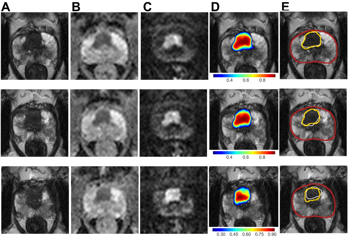
  • U-Net maintained similar diagnostic performance compared to radiological assessment of PI-RADS ≥ 4 when applied in a simulated clinical deployment.
  • Application of our proposed prospective dynamic calibration method successfully adjusted U-Net performance within acceptable limits of the PI-RADS reference over time, while not being limited to PI-RADS as a reference.
  • Simultaneous detection by U-Net and radiological assessment significantly improved the positive predictive value on a per-patient and per-lesion basis, while the negative predictive value remained unchanged.

Article: Simulated clinical deployment of fully automatic deep learning for clinical prostate MRI assessment

Authors: Patrick Schelb, Xianfeng Wang, Jan Philipp Radtke, Manuel Wiesenfarth, Philipp Kickingereder, Albrecht Stenzinger, Markus Hohenfellner, Heinz-Peter Schlemmer, Klaus H. Maier-Hein & David Bonekamp

WRITTEN BY

  • David Bonekamp

    Division of Radiology, Germany Cancer Research Center (DKFZ), Heidelberg, Germany

Latest posts

Become A Member Today!

You will have access to a wide range of benefits that can help you advance your career and stay up-to-date with the latest developments in the field of radiology. These benefits include access to educational resources, networking opportunities with other professionals in the field, opportunities to participate in research projects and clinical trials, and access to the latest technologies and techniques. 

Check out our different membership options.

If you don’t find a fitting membership send us an email here.

Membership

for radiologists, radiology residents, professionals of allied sciences (including radiographers/radiological technologists, nuclear medicine physicians, medical physicists, and data scientists) & professionals of allied sciences in training residing within the boundaries of Europe

  • Reduced registration fees for ECR 1
  • Reduced fees for the European School of Radiology (ESOR) 2
  • Exclusive option to participate in the European Diploma. 3
  • Free electronic access to the journal European Radiology 4
  • Content e-mails for all ESR journals
  • Updates on offers & events through our newsletters
  • Exclusive access to the ESR feed in Juisci

€ 11 /year

Yes! That is less than €1 per month.

Free membership

for radiologists, radiology residents or professionals of allied sciences engaged in practice, teaching or research residing outside Europe as well as individual qualified professionals with an interest in radiology and medical imaging who do not fulfil individual or all requirements for any other ESR membership category & former full members who have retired from all clinical practice
  • Reduced registration fees for ECR 1
  • Free electronic access to the journal European Radiology
  • Content e-mails for all 3 ESR journals 4
  • Updates on offers & events through our newsletters
  • Exclusive access to the ESR feed in Juisci

€ 0

The best things in life are free.

ESR Friends

For students, company representatives or hospital managers etc.

  • Content e-mails for all 3 ESR journals 4
  • Updates on offers & events through our newsletters

€ 0

Friendship doesn’t cost a thing.

The membership type best fitting for you will be selected automatically during the application process.

Footnotes:

01

Reduced registration fees for ECR 2024:
Provided that ESR 2023 membership is activated and approved by August 31, 2023.

Reduced registration fees for ECR 2025:
Provided that ESR 2024 membership is activated and approved by August 31, 2024.

02
Not all activities included
03
Examination based on the ESR European Training Curriculum (radiologists or radiology residents).
04
European Radiology, Insights into Imaging, European Radiology Experimental.