Standardised lesion segmentation for imaging biomarker quantitation

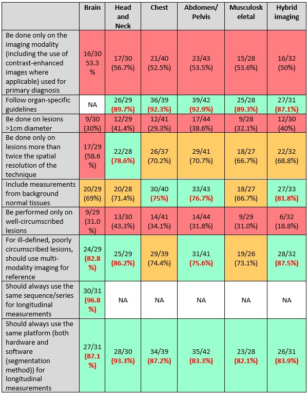
Extraction of quantitative data from images requires selecting the regions-of-interest from which this data is to be extracted. This process of region-of-interest selection (or segmentation), whether it be manual or automated, crucially determines the values of the extracted biomarkers. Harmonisation of the segmentation process itself is therefore central to the standardisation of imaging biomarkers. We undertook a Delphi process amongst >50 multidisciplinary experts to achieve a consensus for segmentation practice on CT, MRI and PET-CT images. Items with a >75% agreement were put forward as a recommendation. We concluded that while operator training with appropriate refreshment was absolutely essential in clinical trials and clinical research, board certification of operators was not required. We also recommend system certification and specification of the images to be used, with specified thresholds for signal-to-noise and contrast-to-noise ratios, and study-specific tumour-to background ratios in PET. We strongly recommend SOPs for quantitative imaging with window-level ranges specified on an organ basis. We believe that while multiple reconstruction methods can be used, along with a mixture of CE marked and research software for segmentation, validation of the methods is mandatory, with human observer reference standards essential for both manual and automated segmentation processes. For poorly defined lesions we recommend using multi-modality imaging as a reference. We also recommend that measurements from background normal tissues should be made and longitudinal measurements strictly use the same hardware and software platforms.

Key points

  • System certification and specification of image characteristics is recommended for segmentation.
  • Both CE marked and verified research tools are allowable for segmentation.
  • Operator training and refreshment are mandatory for segmentation in clinical trials and research.
  • Pixel numbers within lesion segmented and post-reconstruction algorithms used need reporting.
  • Board certification of operators and frequency of re-training need not be mandated.

Article: Standardised lesion segmentation for imaging biomarker quantitation: a consensus recommendation from ESR and EORTC

Authors: Nandita M. deSouza, Aad van der Lugt, Christophe M. Deroose, Angel Alberich-Bayarri, Luc Bidaut, Laure Fournier, Lena Costaridou, Daniela E. Oprea-Lager, Elmar Kotter, Marion Smits, Marius E. Mayerhoefer, Ronald Boellaard, Anna Caroli, Lioe-Fee de Geus-Oei, Wolfgang G. Kunz, Edwin H. Oei, Frederic Lecouvet, Manuela Franca, Christian Loewe, Egesta Lopci, Caroline Caramella, Anders Persson, Xavier Golay, Marc Dewey, James P. B. O’Connor, Pim deGraaf, Sergios Gatidis, Gudrun Zahlmann, European Society of Radiology & European Organisation for Research and Treatment of Cancer

WRITTEN BY

  • Nandita deSouza

    Division of Radiotherapy and Imaging, The Institute of Cancer Research and Royal Marsden NHS Foundation Trust, London, UK

Latest posts

Become A Member Today!

You will have access to a wide range of benefits that can help you advance your career and stay up-to-date with the latest developments in the field of radiology. These benefits include access to educational resources, networking opportunities with other professionals in the field, opportunities to participate in research projects and clinical trials, and access to the latest technologies and techniques. 

Check out our different membership options.

If you don’t find a fitting membership send us an email here.

Membership

for radiologists, radiology residents, professionals of allied sciences (including radiographers/radiological technologists, nuclear medicine physicians, medical physicists, and data scientists) & professionals of allied sciences in training residing within the boundaries of Europe

  • Reduced registration fees for ECR 1
  • Reduced fees for the European School of Radiology (ESOR) 2
  • Exclusive option to participate in the European Diploma. 3
  • Free electronic access to the journal European Radiology 4
  • Content e-mails for all ESR journals
  • Updates on offers & events through our newsletters
  • Exclusive access to the ESR feed in Juisci

€ 11 /year

Yes! That is less than €1 per month.

Free membership

for radiologists, radiology residents or professionals of allied sciences engaged in practice, teaching or research residing outside Europe as well as individual qualified professionals with an interest in radiology and medical imaging who do not fulfil individual or all requirements for any other ESR membership category & former full members who have retired from all clinical practice
  • Reduced registration fees for ECR 1
  • Free electronic access to the journal European Radiology
  • Content e-mails for all 3 ESR journals 4
  • Updates on offers & events through our newsletters
  • Exclusive access to the ESR feed in Juisci

€ 0

The best things in life are free.

ESR Friends

For students, company representatives or hospital managers etc.

  • Content e-mails for all 3 ESR journals 4
  • Updates on offers & events through our newsletters

€ 0

Friendship doesn’t cost a thing.

The membership type best fitting for you will be selected automatically during the application process.

Footnotes:

01

Reduced registration fees for ECR 2024:
Provided that ESR 2023 membership is activated and approved by August 31, 2023.

Reduced registration fees for ECR 2025:
Provided that ESR 2024 membership is activated and approved by August 31, 2024.

02
Not all activities included
03
Examination based on the ESR European Training Curriculum (radiologists or radiology residents).
04
European Radiology, Insights into Imaging, European Radiology Experimental.