The role of generative adversarial networks in brain MRI

Magnetic Resonance Imaging (MRI) is a widely used medical imaging technology that is non-intrusive and considered safe for humans and can generate different modalities of an image, as well as provide valuable insights into a specific disease. The frequent sequences of MRI are T1-weighted and T2- weighted scans. The popularity of artificial intelligence (AI) for brain MRI is on the rise with many recent developments published in the literature. Typically, the performance of AI for brain MRI can improve if enough data is made available. Generative Adversarial Networks (GANs) showed great potential in generating synthetic MRI data that can capture the distribution of real MRI. Furthermore, GANs are also popular for segmentation, noise removal, and super-resolution of brain MRI images.

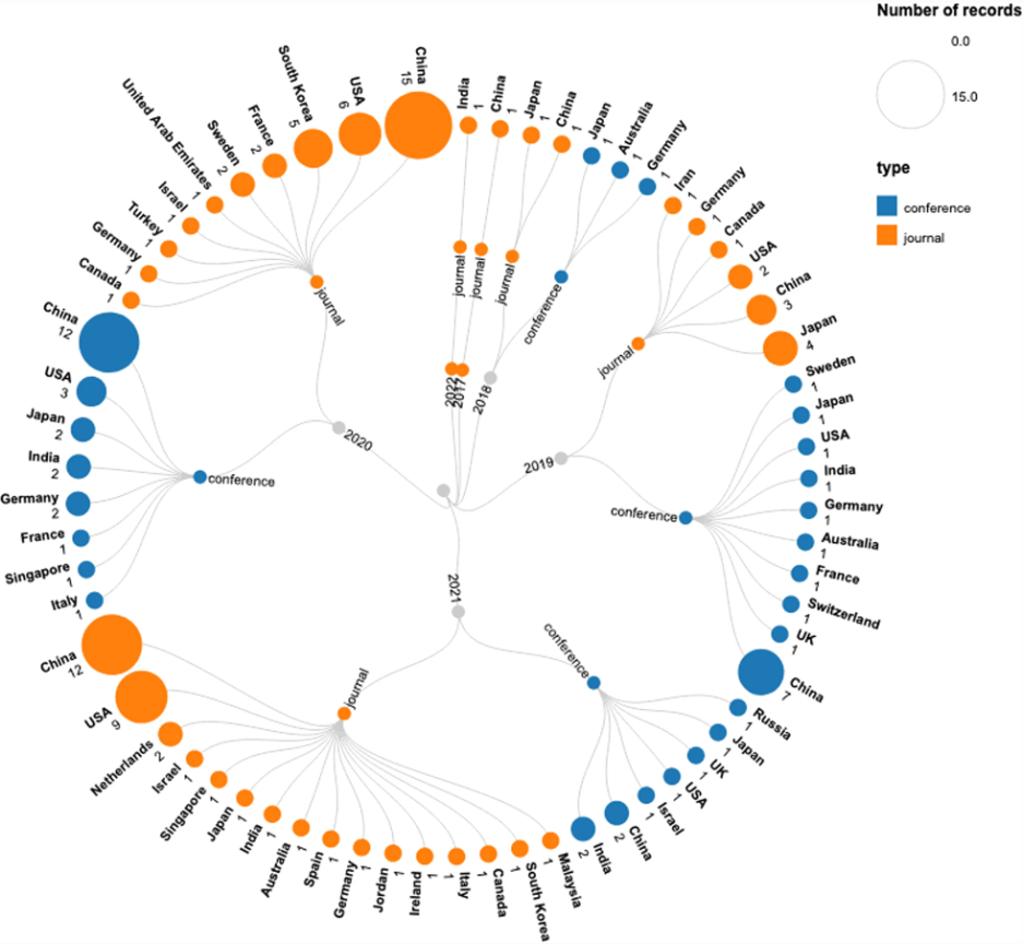
This review explores how GANs methods have been used on brain MRI data, as reported in the literature. The review describes the different applications of GANs for brain MRI, presents the most commonly used GANs architectures, and summarizes the publicly available brain MRI datasets for advancing research and development of GANs-based approaches; The review includes 139 published studies. A careful analysis of the studies reveals that the most common use case of GANs is the synthesis of brain MRI images for data augmentation. GANs have also been used to segment brain tumors and translate healthy images to diseased images or CT to MRI and vice-versa. The included studies showed that GANs could enhance the performance of AI methods used on brain MRI imaging data. However, more efforts are needed to transform the GANs-based methods in clinical applications.

The following research questions related to the role of the GANs-based method in brain MRI were considered for this review:
1. What were the typical applications of GANs proposed for brain MRI?
2. Which architectures of GANs are most commonly applied for brain MRI?
3. What was the purpose of using GAN in brain MRI?
4. What were the most commonly used datasets for brain MRI?
5. How many datasets were publicly accessible?
6. What evaluation matrices were used for the validation of the model?

We believe that this study will be helpful for researchers and professionals in the medical imaging and healthcare domain who are considering using GANs methods for the diagnosis and prognosis of brain tumors from MRI images. The review also lists publicly available brain MRI datasets that will be helpful for AI researchers to develop advanced research methods.

Key points

  • This article aims to provide a comprehensive review on the applications of generative adversarial networks (GANs) in brain MRI.
  • The specific focus of this education review is on brain MRI.
  • It covers a large number of studies on GANs in brain MRI and the most recently published studies on brain MRI.

Article: The role of generative adversarial networks in brain MRI: a scoping review

Authors: Hazrat Ali, Md. Rafiul Biswas, Farida Mohsin, Uzair Shah, Asma Alamgir, Osama Mousa & Zubair Shah

WRITTEN BY

  • Hazrat Ali

    Dr. Hazrat Ali is a researcher in AI for healthcare at the College of Science and Engineering, Hamad Bin Khalifa University, Qatar. His research interests lie in deep learning and medical imaging. He is a senior member of IEEE and an Associate Editor at IEEE Access. He was selected as a young researcher at the 5th Heidelberg Laureate Forum in Heidelberg, Germany and is the recipient of the HEC Scholarship, the Top 10 research pitch award by the University of Queensland, Australia, IEEE Student Travel Award, the IBRO grant, the TERENA/CISCO Travel grant, and the Erasmus Mundus STRoNGTiES research grant.

  • Zubair Shah

    Dr. Zubair Shah is an Assistant Professor at the Division of ICT, College of Science and Engineering, Hamad Bin Khalifa University, Qatar. He was a Research Fellow from 2017-2019 at the Australian Institute of Health Innovation, Macquarie University, Australia. His expertise is in the field of artificial intelligence and big data analytics, and their application to health informatics. His research is focused on health informatics, particularly in relation to public health, using social media data (e.g., Twitter) and news sources to identify patterns indicative of population-level health. He has published his work in various A-tier international journals and conferences.

Latest posts

Become A Member Today!

You will have access to a wide range of benefits that can help you advance your career and stay up-to-date with the latest developments in the field of radiology. These benefits include access to educational resources, networking opportunities with other professionals in the field, opportunities to participate in research projects and clinical trials, and access to the latest technologies and techniques. 

Check out our different membership options.

If you don’t find a fitting membership send us an email here.

Membership

for radiologists, radiology residents, professionals of allied sciences (including radiographers/radiological technologists, nuclear medicine physicians, medical physicists, and data scientists) & professionals of allied sciences in training residing within the boundaries of Europe

  • Reduced registration fees for ECR 1
  • Reduced fees for the European School of Radiology (ESOR) 2
  • Option to participate in the European Diploma. 3
  • Free electronic access to the journal European Radiology 
  • Content e-mails for all ESR journals4
  • Updates on offers & events through our newsletters
  • Exclusive access to the ESR feed in Juisci

€ 11 /year

Yes! That is less than €1 per month.

Free membership

for radiologists, radiology residents or professionals of allied sciences engaged in practice, teaching or research residing outside Europe as well as individual qualified professionals with an interest in radiology and medical imaging who do not fulfil individual or all requirements for any other ESR membership category & former full members who have retired from all clinical practice
  • Reduced registration fees for ECR 1
  • Option to participate in the European Diploma. 3
  • Free electronic access to the journal European Radiology
  • Content e-mails for all ESR journals 4
  • Updates on offers & events through our newsletters
  • Exclusive access to the ESR feed in Juisci

€ 0

The best things in life are free.

ESR Friends

For students, company representatives or hospital managers etc.

  • Content e-mails for all 3 ESR journals 4
  • Updates on offers & events through our newsletters

€ 0

Friendship doesn’t cost a thing.

The membership type best fitting for you will be selected automatically during the application process.

Footnotes:

01

Reduced registration fees for ECR 2025:
Provided that ESR 2024 membership is activated and approved by August 31, 2024.

02
Not all activities included
03
Examination based on the ESR European Training Curriculum (radiologists or radiology residents).
04
European Radiology, Insights into Imaging, European Radiology Experimental.