


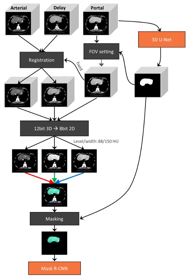
Of late, deep learning-based algorithms have been successfully applied to various medical imaging modalities, ranging from chest radiographs to head CT scans. Compared to other body parts, there is a paucity of data regarding the application of deep learning-based algorithms in the liver. This can be attributed to the following reasons: First, unlike other body parts usually relying on single-phase images, analyzing multiple-phase images of CT or MRI is essential in order to characterize focal hepatic lesions, which requires integration of the complex information; second, the complex vascular anatomy of the liver and its adjacent organs may mimic abnormalities in the liver.
To overcome such obstacles, we developed and evaluated a deep learning-based model to detect hepatic malignancies based on multichannel integration and automatic segmentation of the liver using Mask R-CNN. Our model showed high (84.8%) sensitivity with less than 5 false positives per CT scan on the test set. We believe that our study sheds light on the application of deep learning-based algorithms to detect primary hepatic tumors which have continued to increase in their incidences worldwide.
Key points
- Image processing, including multichannel integration of multiphase CT and automatic liver segmentation, enabled the application of a deep learning-based model to detect primary hepatic malignancy.
- Our model exhibited a sensitivity of 84.8% with a false-positive rate of 4.80 per CT scan.
Authors: Dong Wook Kim, Gaeun Lee, So Yeon Kim, Geunhwi Ahn, June-Goo Lee, Seung Soo Lee, Kyung Won Kim, Seong Ho Park, Yoon Jin Lee & Namkug Kim


