Using machine learning to predict cervical lymph node metastasis on dual-energy CT

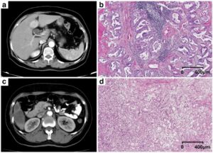
In this article, we extracted “hand-crafted” radiomic features from dual-energy CT (DECT) virtual monochromatic images (VMIs) reconstructed at different energies and used machine learning to construct prediction models that use the radiomic features of head and neck squamous cell carcinoma (HNSCC) to predict associated nodal metastases. This proof of concept study demonstrated that (1) HNSCC radiomic features can predict associated nodal metastases, including potentially micro-metastases (i.e. positive N status), in otherwise radiologically normal nodes, and (2) model performance improved substantially when multi-energy radiomic features were used compared to models constructed using radiomic features extracted from 65 keV VMIs alone, typically considered similar to a single energy CT. The question addressed is particularly clinically relevant and meant to provide the basis for an artificial (or augmented) intelligence (AI) clinical assistant tool to complement expert radiologist assessments. If validated in larger studies, this has the potential to reduce unnecessary neck dissections in HNSCC patients. The next steps would be to use deep extracted features from tumors, lymph nodes, and other routinely available clinical information, to validate and, perhaps, further enhance model performance and bring this closer to an implementable AI clinical assistant tool. The other key observation is the significant advantage conferred by analysis of DECT data. There are currently multiple clinical applications of DECT, but enhanced radiomic and AI applications, and the ability to create more robust non-invasive biomarkers for precision oncology, could represent a new horizon for this technology, with potential applications far beyond the specific model investigated in this study.

Key points

  • Texture features of HNSCC tumor are predictive of nodal status.
  • Multi-energy texture analysis is superior to analysis of datasets at a single energy.
  • Dual-energy CT texture analysis with machine learning can enhance noninvasive diagnostic tumor evaluation.

Article: Head and neck squamous cell carcinoma: prediction of cervical lymph node metastasis by dual-energy CT texture analysis with machine learning

Authors: Reza Forghani, Avishek Chatterjee, Caroline Reinhold, Almudena Pérez-Lara, Griselda Romero-Sanchez, Yoshiko Ueno, Maryam Bayat, James W. M. Alexander, Lynda Kadi, Jeffrey Chankowsky, Jan Seuntjens, Behzad Forghani

WRITTEN BY

Latest posts

Become A Member Today!

You will have access to a wide range of benefits that can help you advance your career and stay up-to-date with the latest developments in the field of radiology. These benefits include access to educational resources, networking opportunities with other professionals in the field, opportunities to participate in research projects and clinical trials, and access to the latest technologies and techniques. 

Check out our different membership options.

If you don’t find a fitting membership send us an email here.

Membership

for radiologists, radiology residents, professionals of allied sciences (including radiographers/radiological technologists, nuclear medicine physicians, medical physicists, and data scientists) & professionals of allied sciences in training residing within the boundaries of Europe

  • Reduced registration fees for ECR 1
  • Reduced fees for the European School of Radiology (ESOR) 2
  • Option to participate in the European Diploma. 3
  • Free electronic access to the journal European Radiology 
  • Content e-mails for all ESR journals4
  • Updates on offers & events through our newsletters
  • Exclusive access to the ESR feed in Juisci

€ 11 /year

Yes! That is less than €1 per month.

Free membership

for radiologists, radiology residents or professionals of allied sciences engaged in practice, teaching or research residing outside Europe as well as individual qualified professionals with an interest in radiology and medical imaging who do not fulfil individual or all requirements for any other ESR membership category & former full members who have retired from all clinical practice
  • Reduced registration fees for ECR 1
  • Option to participate in the European Diploma. 3
  • Free electronic access to the journal European Radiology
  • Content e-mails for all ESR journals 4
  • Updates on offers & events through our newsletters
  • Exclusive access to the ESR feed in Juisci

€ 0

The best things in life are free.

ESR Friends

For students, company representatives or hospital managers etc.

  • Content e-mails for all 3 ESR journals 4
  • Updates on offers & events through our newsletters

€ 0

Friendship doesn’t cost a thing.

The membership type best fitting for you will be selected automatically during the application process.

Footnotes:

01

Reduced registration fees for ECR 2025:
Provided that ESR 2024 membership is activated and approved by August 31, 2024.

02
Not all activities included
03
Examination based on the ESR European Training Curriculum (radiologists or radiology residents).
04
European Radiology, Insights into Imaging, European Radiology Experimental.