machine learning

Welcome to the blog on Artificial Intelligence of
the European Society of Radiology

This blog aims at bringing educational and critical perspectives on AI to readers. It should help imaging professionals to learn and keep up to date with the technologies being developed in this rapidly evolving field.

Most used hashtags:

Latest posts

Coronary CT angiographic detection of in-stent restenosis via deep learning reconstruction

This feasibility study used deep learning reconstruction, Precise IQ Engine (PIQE), to quantify stent strut thickness and lumen vessel diameter, subsequently comparing it with values obtained using conventional reconstruction methods. The authors conducting this study examined 166 stents in 85 consecutive patients who had undergone CT and invasive coronary angiography (ICA) within 3 months of each other. The results demonstrated

Read More →

Radiomics-based prediction of FIGO grade for placenta accreta spectrum

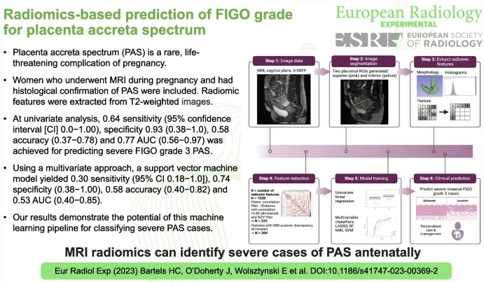
Placenta Accreta Spectrum (PAS) is a serious, life-threatening pregnancy complication. Although rare, as more women are giving birth by Caesarean section, PAS is becoming more common. The most important factor in improving outcomes for mothers and babies is the detection of PAS during pregnancy to ensure the appropriate multi-disciplinary team care is implemented for the pregnancy and birth. However, up

Read More →

Self-reporting with checklists in AI research on medical imaging

This study aimed to evaluate the usage of the Checklist for Artificial Intelligence in Medical Imaging (CLAIM), a well-known and widely adopted checklist in the radiological community, for self-reporting through a systematic analysis of its citations. The authors used three databases (Google Scholar, Web of Science, and Scopus) and identified nearly 400 unique citations across 118 papers, of which only

Read More →

Are deep models in radiomics performing better than generic models?

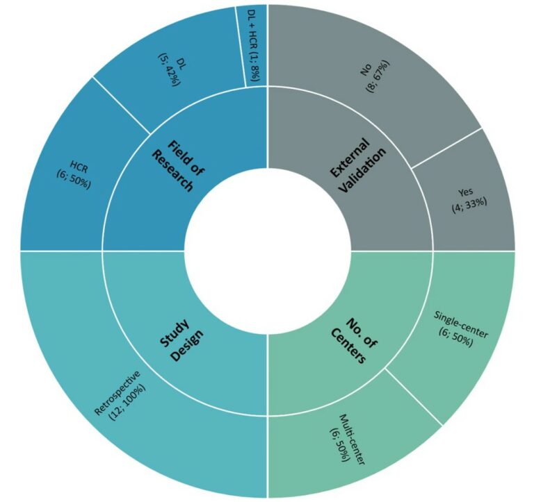
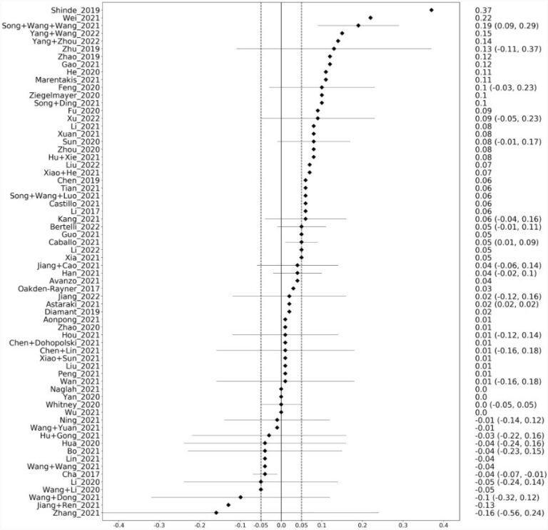
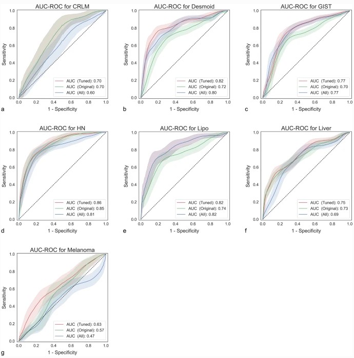
In radiomics, deep learning methods are increasingly used because they promise higher predictive performance than models based on generic, hand-crafted features. However, sample sizes in radiomics are often small, and it is known that the performance of deep learning models is often critically dependent on sample size. Therefore, it is unclear whether deep models can outperform generic models. In our

Read More →

Quo vadis Radiomics? Bibliometric analysis of 10-year Radiomics journey

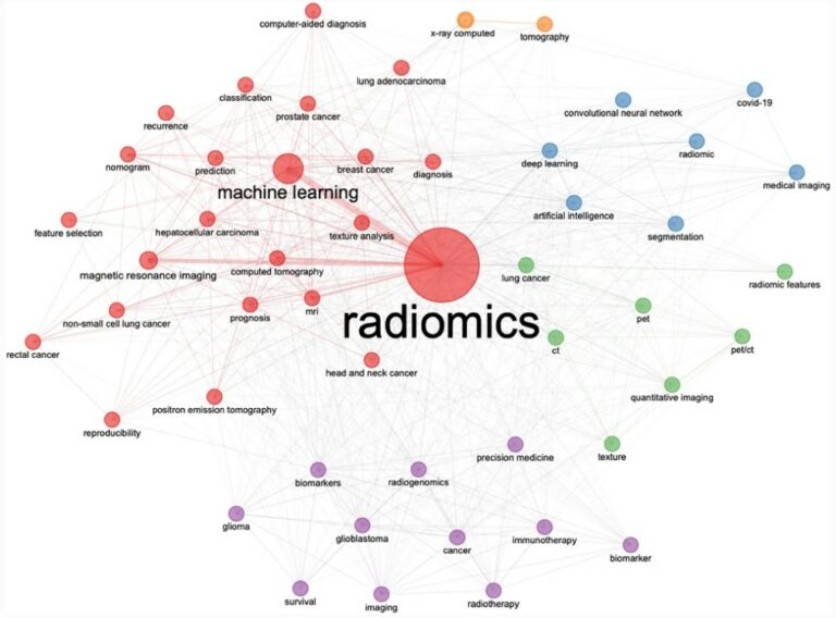
This study conducted a bibliometric analysis of radiomics ten years after the first work became available in March 2012. Throughout the analysis, the authors identified over 5,500 articles from almost 17,000 authors from over 900 different sources, highlighting developments within radiomics, its real-world applications, and tangible and intangible benefits. Key points ML-based bibliometric analysis is fundamental to detect unknown pattern

Read More →

A novel AI model to distinguish benign from malignant ovarian tumors

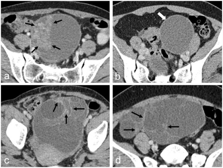
The authors of this study developed a CT-based artificial intelligence model with the ability to differentiate between benign and malignant ovarian tumors, showing high accuracy and specificity. In coordination with less-experienced radiologists, the model helped in the performance of ovarian tumor assessment, with applications to provide better therapeutic strategies for patients with ovarian tumors. Key points CT-based radiomics and deep

Read More →

Automated vetting of radiology referrals: exploring NLP and ML approaches

As computed tomography (CT) sees an increase in utilization, inappropriate imaging has been seen as a significant concern; however, manual justification audits of radiology referrals are extremely time-consuming and carry a heavy financial burden. Therefore, the authors of this study aimed to retrospectively audit the justification of brain CT referrals by using natural language processing (NLP) and traditional machine learning

Read More →

T2-weighted MRI-based radiomics discriminati between benign and borderline epithelial ovarian tumors

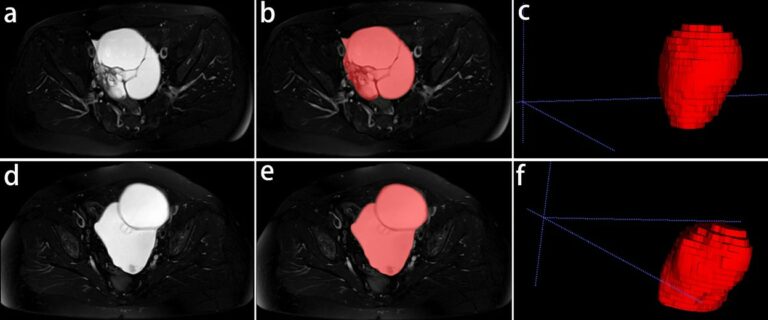
The authors of this study investigated whether radiomics based on T2-weighted MRI was able to discriminate between benign and borderline epithelial ovarian tumors (EOTs) preoperatively. They were able to show that it can provide critical diagnostic information in the discrimination between benign and borderline EOTs, showing that there is potential to aid personalized treatment options. Key points T2-weighted MRI-based radiomics

Read More →

Tasks for AI in prostate MRI

The authors of this narrative review aimed to introduce quality metrics for emerging artificial intelligence (AI) papers, such as the Checklist for Artificial Intelligence in Medical Imaging (CLAIM) and Field-Weighted Citation Impact (FWCI). Furthermore, the study dives into some of the top AI models for segmentation, detection, and classification, while concluding that prospective studies with multi-center design will need to

Read More →

Utilizing CLAIM to adapt the increasing trend of deep learning application in radiomics

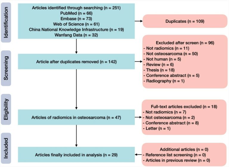
The aim of this study was to provide an updated systematic review of radiomics in osteosarcoma, utilizing various databases such as PubMed, Embase, China National Knowledge Infrastructure, and more. Articles found in these databases were assessed by Radiomics Quality Score (RQS), Transparent Reporting of a multivariable prediction model for Individual Prognosis Or Diagnosis (TRIPOD) statement, Checklist for Artificial Intelligence in

Read More →

Coronary CT angiographic detection of in-stent restenosis via deep learning reconstruction

This feasibility study used deep learning reconstruction, Precise IQ Engine (PIQE), to quantify stent strut thickness and lumen vessel diameter, subsequently comparing it with values obtained using conventional reconstruction methods. The authors conducting this study examined 166 stents in 85 consecutive patients who had undergone CT and invasive coronary angiography (ICA) within 3 months of each other. The results demonstrated

Read More →

Radiomics-based prediction of FIGO grade for placenta accreta spectrum

Placenta Accreta Spectrum (PAS) is a serious, life-threatening pregnancy complication. Although rare, as more women are giving birth by Caesarean section, PAS is becoming more common. The most important factor in improving outcomes for mothers and babies is the detection of PAS during pregnancy to ensure the appropriate multi-disciplinary team care is implemented for the pregnancy and birth. However, up

Read More →

Self-reporting with checklists in AI research on medical imaging

This study aimed to evaluate the usage of the Checklist for Artificial Intelligence in Medical Imaging (CLAIM), a well-known and widely adopted checklist in the radiological community, for self-reporting through a systematic analysis of its citations. The authors used three databases (Google Scholar, Web of Science, and Scopus) and identified nearly 400 unique citations across 118 papers, of which only

Read More →

Are deep models in radiomics performing better than generic models?

In radiomics, deep learning methods are increasingly used because they promise higher predictive performance than models based on generic, hand-crafted features. However, sample sizes in radiomics are often small, and it is known that the performance of deep learning models is often critically dependent on sample size. Therefore, it is unclear whether deep models can outperform generic models. In our

Read More →

Quo vadis Radiomics? Bibliometric analysis of 10-year Radiomics journey

This study conducted a bibliometric analysis of radiomics ten years after the first work became available in March 2012. Throughout the analysis, the authors identified over 5,500 articles from almost 17,000 authors from over 900 different sources, highlighting developments within radiomics, its real-world applications, and tangible and intangible benefits. Key points ML-based bibliometric analysis is fundamental to detect unknown pattern

Read More →

A novel AI model to distinguish benign from malignant ovarian tumors

The authors of this study developed a CT-based artificial intelligence model with the ability to differentiate between benign and malignant ovarian tumors, showing high accuracy and specificity. In coordination with less-experienced radiologists, the model helped in the performance of ovarian tumor assessment, with applications to provide better therapeutic strategies for patients with ovarian tumors. Key points CT-based radiomics and deep

Read More →

Automated vetting of radiology referrals: exploring NLP and ML approaches

As computed tomography (CT) sees an increase in utilization, inappropriate imaging has been seen as a significant concern; however, manual justification audits of radiology referrals are extremely time-consuming and carry a heavy financial burden. Therefore, the authors of this study aimed to retrospectively audit the justification of brain CT referrals by using natural language processing (NLP) and traditional machine learning

Read More →

T2-weighted MRI-based radiomics discriminati between benign and borderline epithelial ovarian tumors

The authors of this study investigated whether radiomics based on T2-weighted MRI was able to discriminate between benign and borderline epithelial ovarian tumors (EOTs) preoperatively. They were able to show that it can provide critical diagnostic information in the discrimination between benign and borderline EOTs, showing that there is potential to aid personalized treatment options. Key points T2-weighted MRI-based radiomics

Read More →

Tasks for AI in prostate MRI

The authors of this narrative review aimed to introduce quality metrics for emerging artificial intelligence (AI) papers, such as the Checklist for Artificial Intelligence in Medical Imaging (CLAIM) and Field-Weighted Citation Impact (FWCI). Furthermore, the study dives into some of the top AI models for segmentation, detection, and classification, while concluding that prospective studies with multi-center design will need to

Read More →

Utilizing CLAIM to adapt the increasing trend of deep learning application in radiomics

The aim of this study was to provide an updated systematic review of radiomics in osteosarcoma, utilizing various databases such as PubMed, Embase, China National Knowledge Infrastructure, and more. Articles found in these databases were assessed by Radiomics Quality Score (RQS), Transparent Reporting of a multivariable prediction model for Individual Prognosis Or Diagnosis (TRIPOD) statement, Checklist for Artificial Intelligence in

Read More →

Become A Member Today!

You will have access to a wide range of benefits that can help you advance your career and stay up-to-date with the latest developments in the field of radiology. These benefits include access to educational resources, networking opportunities with other professionals in the field, opportunities to participate in research projects and clinical trials, and access to the latest technologies and techniques. 

Check out our different membership options.

If you don’t find a fitting membership send us an email here.

Membership

for radiologists, radiology residents, professionals of allied sciences (including radiographers/radiological technologists, nuclear medicine physicians, medical physicists, and data scientists) & professionals of allied sciences in training residing within the boundaries of Europe

  • Reduced registration fees for ECR 1
  • Reduced fees for the European School of Radiology (ESOR) 2
  • Option to participate in the European Diploma. 3
  • Free electronic access to the journal European Radiology 
  • Content e-mails for all ESR journals4
  • Updates on offers & events through our newsletters
  • Exclusive access to the ESR feed in Juisci

€ 11 /year

Yes! That is less than €1 per month.

Free membership

for radiologists, radiology residents or professionals of allied sciences engaged in practice, teaching or research residing outside Europe as well as individual qualified professionals with an interest in radiology and medical imaging who do not fulfil individual or all requirements for any other ESR membership category & former full members who have retired from all clinical practice
  • Reduced registration fees for ECR 1
  • Option to participate in the European Diploma. 3
  • Free electronic access to the journal European Radiology
  • Content e-mails for all ESR journals 4
  • Updates on offers & events through our newsletters
  • Exclusive access to the ESR feed in Juisci

€ 0

The best things in life are free.

ESR Friends

For students, company representatives or hospital managers etc.

  • Content e-mails for all 3 ESR journals 4
  • Updates on offers & events through our newsletters

€ 0

Friendship doesn’t cost a thing.

The membership type best fitting for you will be selected automatically during the application process.

Footnotes:

01

Reduced registration fees for ECR 2025:
Provided that ESR 2024 membership is activated and approved by August 31, 2024.

02
Not all activities included
03
Examination based on the ESR European Training Curriculum (radiologists or radiology residents).
04
European Radiology, Insights into Imaging, European Radiology Experimental.