tomography

Welcome to the blog on Artificial Intelligence of
the European Society of Radiology

This blog aims at bringing educational and critical perspectives on AI to readers. It should help imaging professionals to learn and keep up to date with the technologies being developed in this rapidly evolving field.

Most used hashtags:

Latest posts

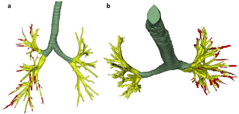
Creating a training set for AI from initial segmentations of airways

An important challenge in the use of artificial intelligence (AI) for medical image segmentation tasks is the lack of high-quality, scan protocol-specific datasets. AI performs best on narrow tasks with homogenous specifications. Thus, pre-trained models may be inadequate for use in centre-specific studies if the scan protocols do not match. For the airway segmentation task in the Imaging in Lifelines

Read More →

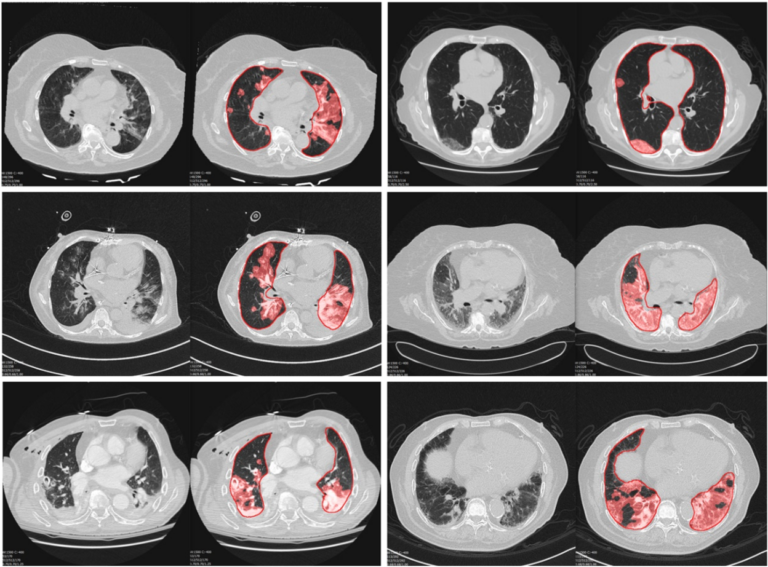
Machine learning automatically detects COVID-19 using chest CTs in a large multicenter cohort

This retrospective, multi-institutional study investigated machine learning classifiers and interpretable models using chest CT for the detection of COVID-19 and to differentiate this from types of pneumonia, interstitial lung disease (ILD), and normal CTs. The study included 2,446 chest CTs from across 16 different institutions and the authors’ method was found to accurately differentiate COVID-19 from other types of pneumonia,

Read More →

A comparison between manual and artificial intelligence–based automatic positioning in CT imaging for COVID-19 patients

The authors of this study analyzed and compared the imaging workflow, radiation dose, and image quality for 127 adult COVID-19 patients who were examined using either the conventional manual positioning (MP) method or an AI-based automatic positioning (AP) method. The patients underwent chest CT scans using the manual positioning (MP group) for the initial scan, followed by an AI-based automatic

Read More →

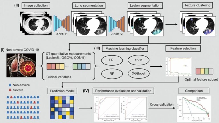
Machine learning based on clinical characteristics and chest CT quantitative measurements for prediction of adverse clinical outcomes in hospitalized patients with COVID-19

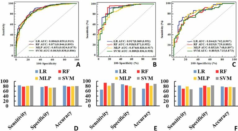
In this study, the authors aimed to develop and validate a machine learning model for the prediction of adverse outcomes in hospitalized patients with COVID-19. They discovered that their findings could be used to facilitate the prediction of adverse outcomes in patients with COVID-19, as well as may allow efficient utilization of medical resources and individualized treatment plans for COVID-19

Read More →

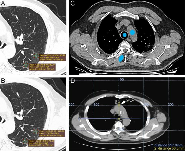
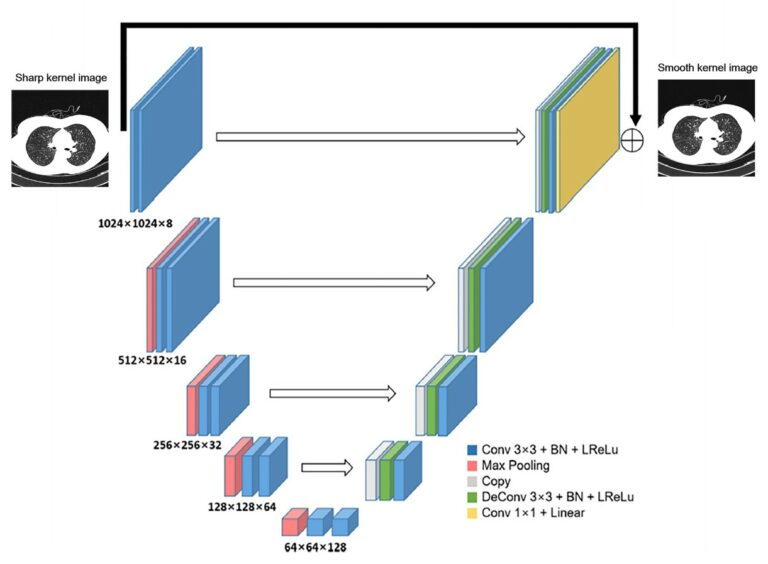
Emphysema quantification using low-dose computed tomography with deep learning-based kernel conversion comparison

This study used a sample of 131 participants who underwent low-dose computed tomography (LDCT) and standard-dose computed tomography (SDCT) to determine the effect of dose reduction and kernel selection on quantifying emphysema. The authors determined that the deep learning-based CT kernel conversation of sharp kernel in LDCT significantly reduced the variation in emphysema quantification. Key points Low-dose computed tomography with

Read More →

Deep learning detection and quantification of pneumothorax in heterogeneous routine chest computed tomography

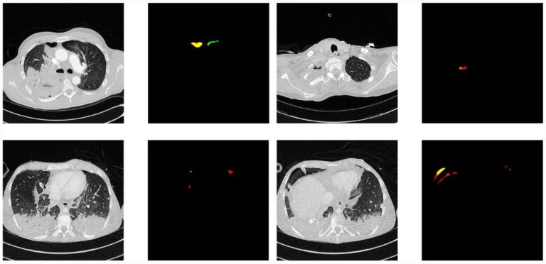
In this study, the authors proposed a deep learning method for the detection and quantification of pneumothorax in heterogeneous routine clinical data, which may facilitate the automated triage of urgent examinations and enable support in the treatment decision. Key points Pneumothorax is an important pathology to be included in applications that are designed to triage urgent imaging examinations. Heterogeneity in

Read More →

Development and validation of machine learning prediction model based on computed tomography angiography–derived hemodynamics for rupture status of intracranial aneurysms: a Chinese multicenter study

Decisions regarding the optimal management of unruptured intracranial aneurysms (UIAs) depend on a comprehensive comparison of the risks between aneurysm rupture and interventional treatment. The accurate prediction for UIA rupture risk is important for clinicians and patients. Our study further proves that the hemodynamic parameters can improve prediction performance for rupture status of UIAs. Moreover, the AUC of model integrating

Read More →

Multiphase CT-based prediction of Child-Pugh classification: a machine learning approach

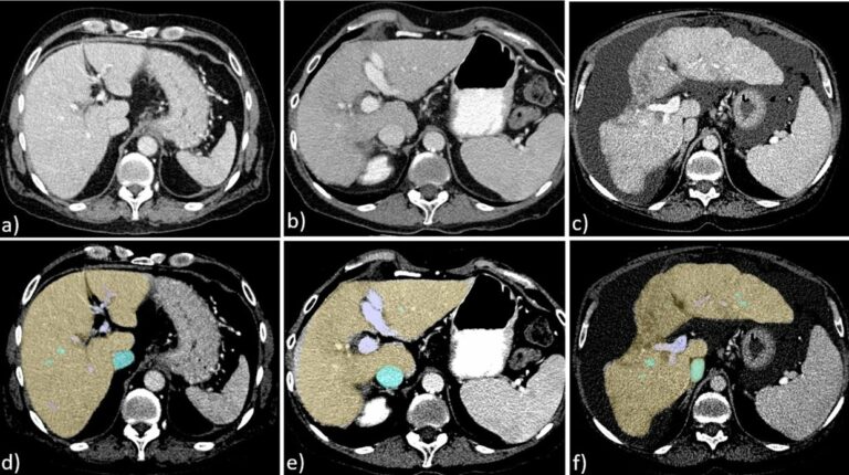
The aim of this study was to evaluate whether machine learning algorithms allow for the prediction of Child-Pugh classification on clinical multiphase computed tomography (CT). The authors found that the performance of convolutional neural networks (CNN) is comparable to that of experienced radiologists in assessing Child-Pugh class based on multiphase abdominal CT. Key points Established machine learning algorithms can predict

Read More →

Radiomics allows for detection of benign and malignant histopathology in patients with metastatic testicular germ cell tumors prior to post-chemotherapy retroperitoneal Iymph node dissection

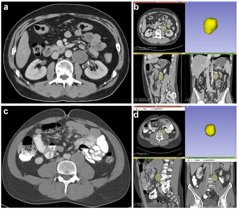
Among solid tumors, testicular germ cell tumors (TGCT) are the most common entity in men below the age of 40, and they are very likely to metastasize. The so-far unsolved clinical problem for metastasized TGCTs is that as many as 40-50% of patients are overtreated by post-chemotherapy lymph node dissection since the resected lymph nodes show a viable tumor in

Read More →

Dual-energy CT–based deep learning radiomics can improve lymph node metastasis risk prediction for gastric cancer

In this study, the authors aimed to build a dual-energy CT (DECT)-based deep learning radiomics nomogram that could be used for lymph node metastasis prediction in gastric cancer. Ultimately, the DECT-based deep learning radiomics nomogram operated well in predicting lymph node metastasis in gastric cancer. Key points This study investigated the value of deep learning dual-energy CT–based radiomics in predicting

Read More →

Creating a training set for AI from initial segmentations of airways

An important challenge in the use of artificial intelligence (AI) for medical image segmentation tasks is the lack of high-quality, scan protocol-specific datasets. AI performs best on narrow tasks with homogenous specifications. Thus, pre-trained models may be inadequate for use in centre-specific studies if the scan protocols do not match. For the airway segmentation task in the Imaging in Lifelines

Read More →

Machine learning automatically detects COVID-19 using chest CTs in a large multicenter cohort

This retrospective, multi-institutional study investigated machine learning classifiers and interpretable models using chest CT for the detection of COVID-19 and to differentiate this from types of pneumonia, interstitial lung disease (ILD), and normal CTs. The study included 2,446 chest CTs from across 16 different institutions and the authors’ method was found to accurately differentiate COVID-19 from other types of pneumonia,

Read More →

A comparison between manual and artificial intelligence–based automatic positioning in CT imaging for COVID-19 patients

The authors of this study analyzed and compared the imaging workflow, radiation dose, and image quality for 127 adult COVID-19 patients who were examined using either the conventional manual positioning (MP) method or an AI-based automatic positioning (AP) method. The patients underwent chest CT scans using the manual positioning (MP group) for the initial scan, followed by an AI-based automatic

Read More →

Machine learning based on clinical characteristics and chest CT quantitative measurements for prediction of adverse clinical outcomes in hospitalized patients with COVID-19

In this study, the authors aimed to develop and validate a machine learning model for the prediction of adverse outcomes in hospitalized patients with COVID-19. They discovered that their findings could be used to facilitate the prediction of adverse outcomes in patients with COVID-19, as well as may allow efficient utilization of medical resources and individualized treatment plans for COVID-19

Read More →

Emphysema quantification using low-dose computed tomography with deep learning-based kernel conversion comparison

This study used a sample of 131 participants who underwent low-dose computed tomography (LDCT) and standard-dose computed tomography (SDCT) to determine the effect of dose reduction and kernel selection on quantifying emphysema. The authors determined that the deep learning-based CT kernel conversation of sharp kernel in LDCT significantly reduced the variation in emphysema quantification. Key points Low-dose computed tomography with

Read More →

Deep learning detection and quantification of pneumothorax in heterogeneous routine chest computed tomography

In this study, the authors proposed a deep learning method for the detection and quantification of pneumothorax in heterogeneous routine clinical data, which may facilitate the automated triage of urgent examinations and enable support in the treatment decision. Key points Pneumothorax is an important pathology to be included in applications that are designed to triage urgent imaging examinations. Heterogeneity in

Read More →

Development and validation of machine learning prediction model based on computed tomography angiography–derived hemodynamics for rupture status of intracranial aneurysms: a Chinese multicenter study

Decisions regarding the optimal management of unruptured intracranial aneurysms (UIAs) depend on a comprehensive comparison of the risks between aneurysm rupture and interventional treatment. The accurate prediction for UIA rupture risk is important for clinicians and patients. Our study further proves that the hemodynamic parameters can improve prediction performance for rupture status of UIAs. Moreover, the AUC of model integrating

Read More →

Multiphase CT-based prediction of Child-Pugh classification: a machine learning approach

The aim of this study was to evaluate whether machine learning algorithms allow for the prediction of Child-Pugh classification on clinical multiphase computed tomography (CT). The authors found that the performance of convolutional neural networks (CNN) is comparable to that of experienced radiologists in assessing Child-Pugh class based on multiphase abdominal CT. Key points Established machine learning algorithms can predict

Read More →

Radiomics allows for detection of benign and malignant histopathology in patients with metastatic testicular germ cell tumors prior to post-chemotherapy retroperitoneal Iymph node dissection

Among solid tumors, testicular germ cell tumors (TGCT) are the most common entity in men below the age of 40, and they are very likely to metastasize. The so-far unsolved clinical problem for metastasized TGCTs is that as many as 40-50% of patients are overtreated by post-chemotherapy lymph node dissection since the resected lymph nodes show a viable tumor in

Read More →

Dual-energy CT–based deep learning radiomics can improve lymph node metastasis risk prediction for gastric cancer

In this study, the authors aimed to build a dual-energy CT (DECT)-based deep learning radiomics nomogram that could be used for lymph node metastasis prediction in gastric cancer. Ultimately, the DECT-based deep learning radiomics nomogram operated well in predicting lymph node metastasis in gastric cancer. Key points This study investigated the value of deep learning dual-energy CT–based radiomics in predicting

Read More →

Become A Member Today!

You will have access to a wide range of benefits that can help you advance your career and stay up-to-date with the latest developments in the field of radiology. These benefits include access to educational resources, networking opportunities with other professionals in the field, opportunities to participate in research projects and clinical trials, and access to the latest technologies and techniques. 

Check out our different membership options.

If you don’t find a fitting membership send us an email here.

Membership

for radiologists, radiology residents, professionals of allied sciences (including radiographers/radiological technologists, nuclear medicine physicians, medical physicists, and data scientists) & professionals of allied sciences in training residing within the boundaries of Europe

  • Reduced registration fees for ECR 1
  • Reduced fees for the European School of Radiology (ESOR) 2
  • Option to participate in the European Diploma. 3
  • Free electronic access to the journal European Radiology 
  • Content e-mails for all ESR journals4
  • Updates on offers & events through our newsletters
  • Exclusive access to the ESR feed in Juisci

€ 11 /year

Yes! That is less than €1 per month.

Free membership

for radiologists, radiology residents or professionals of allied sciences engaged in practice, teaching or research residing outside Europe as well as individual qualified professionals with an interest in radiology and medical imaging who do not fulfil individual or all requirements for any other ESR membership category & former full members who have retired from all clinical practice
  • Reduced registration fees for ECR 1
  • Option to participate in the European Diploma. 3
  • Free electronic access to the journal European Radiology
  • Content e-mails for all ESR journals 4
  • Updates on offers & events through our newsletters
  • Exclusive access to the ESR feed in Juisci

€ 0

The best things in life are free.

ESR Friends

For students, company representatives or hospital managers etc.

  • Content e-mails for all 3 ESR journals 4
  • Updates on offers & events through our newsletters

€ 0

Friendship doesn’t cost a thing.

The membership type best fitting for you will be selected automatically during the application process.

Footnotes:

01

Reduced registration fees for ECR 2025:
Provided that ESR 2024 membership is activated and approved by August 31, 2024.

Reduced registration fees for ECR 2026:
Provided that ESR 2025 membership is activated and approved by August 31, 2025.

02
Not all activities included
03
Examination based on the ESR European Training Curriculum (radiologists or radiology residents).
04
European Radiology, Insights into Imaging, European Radiology Experimental.