radiography

Welcome to the blog on Artificial Intelligence of
the European Society of Radiology

This blog aims at bringing educational and critical perspectives on AI to readers. It should help imaging professionals to learn and keep up to date with the technologies being developed in this rapidly evolving field.

Most used hashtags:

Latest posts

A deep learning algorithm for VHD diagnosis and evaluation

This study aims to develop and validate a deep learning-based automatic chest radiograph (CXR) cardiovascular border (CB) analysis algorithm (CB_auto) in order to diagnose and quantitatively evaluate valvular heart disease (VHD). The authors found that the CB_auto system, in coordination with the deep learning algorithm, provided highly reliable CB measurements, which, in turn, can be useful, not long daily clinical

Read More →

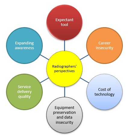
Artificial intelligence in medical imaging practice in Africa: a qualitative content analysis study of radiographers’ perspectives

The aim of this study was to qualitatively explore the perception of radiographers in relation to the integration and acceptance of artificial intelligence (AI) in medical imaging practice on the continent of Africa. Participants of this study consisted solely of radiographers working in Africa between March and August 2020. The study demonstrated a positive outlook regarding AI in relation to

Read More →

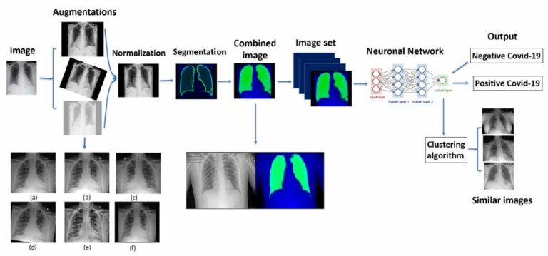
COVID-19 classification of X-ray images using deep neural networks

The authors of this retrospective study propose a deep learning model for the detection of COVID-19 from chest x-rays (CXRs), as well as a tool for retrieving similar patients according to the model’s results on their CXRs. The data used for training and evaluating this model was collected from inpatients across four different hospitals. The proposed model achieved accuracy of

Read More →

AI-based improvement in lung cancer detection on chest radiographs: results of a multi-reader study in NLST dataset

Our study included 519 screening chest radiographs (CXRs) from 294 patients enrolled in the National Lung Screening Trial (NLST) who either had proven to have lung cancer or did not have lung cancer over the duration of the trial. Five attending radiologists and three radiology residents from South Korea and the U.S. independently assessed all CXRs for the presence of

Read More →

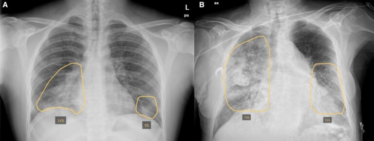
Initial chest radiographs and artificial intelligence (AI) predict clinical outcomes in COVID-19 patients: analysis of 697 Italian patients

The purpose of this retrospective study was to evaluate whether initial chest X-ray (CXR) severity assessed by an AI system may have prognostic utility in patients with COVID-19. The authors determined, through AI- and radiologist-assessed disease severity scores on CXRs obtained on emergency department (ED) presentation, that they were independent and comparable predictors of adverse outcomes in patients with COVID-19.

Read More →

Practical applications of deep learning: classifying the most common categories of plain radiographs in a PACS using a neural network

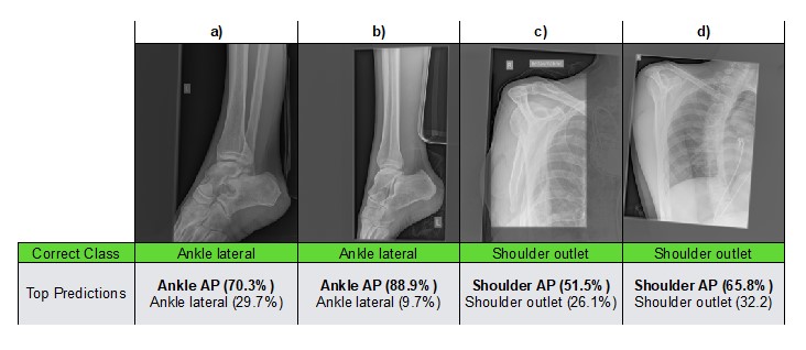
The purpose of this study was to classify the most common types of plain radiography through the use of a neural network and, subsequently, to validate the network’s performance on internal and external data. The authors used data from a single institution when classifying the most common categories of radiographs. This study resulted in the authors determining that it is

Read More →

Deep learning–based automated detection algorithm for active pulmonary tuberculosis on chest radiographs: diagnostic performance in systematic screening of asymptomatic individuals

Chest radiographs (CRs) have long been used as one of the screening tests for pulmonary tuberculosis (TB). However, the interpretation of a large number of CRs is time-consuming and labor-intensive. To overcome this difficulty, we developed the deep-learning-based automated detection (DLAD) for active pulmonary TB detection and performed out-of-sample testing in the consecutively collected 20.135 CRs from 19.686 servicepersons. As

Read More →

Test-retest reproducibility of a deep learning–based automatic detection algorithm for the chest radiograph

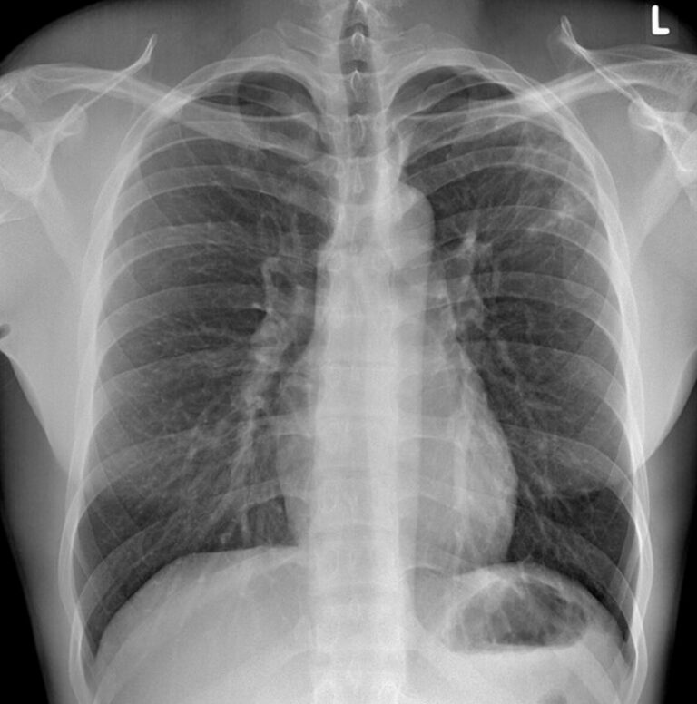
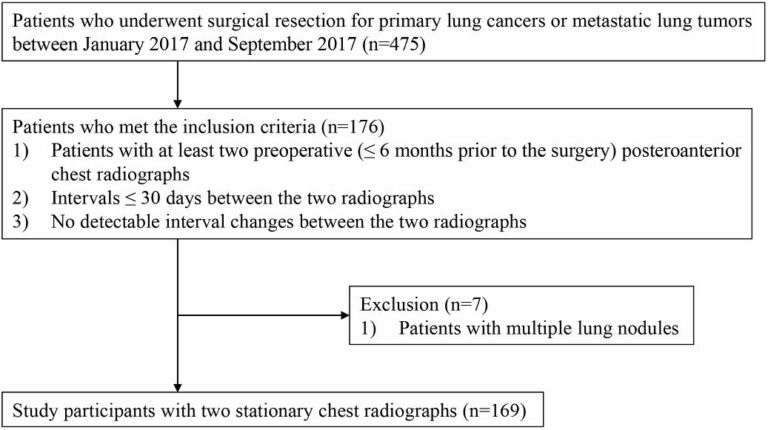
The authors of this retrospective study performed test-retest reproducibility analyses for a deep learning-based automatic detection algorithm (DLAD) using two stationary chest radiographs with short-term intervals, in order to analyze influential factors on test-retest variations. The test, which included patients with pulmonary nodules resected in 2017, showed that DLAD was robust to the test-retest variation. Key points The deep learning–based

Read More →

What the increasing presence of AI means for radiographers

In an age of uncertainty with the arrival of artificial intelligence (AI) tools and technologies in the healthcare field, many in the industry question how the addition of AI will impact their careers. One particular area is not immune to these changes: radiography. We spoke with Dr. Nick Woznitza, a reporting radiographer at Homerton University Hospital and a clinical academic

Read More →

A deep learning algorithm for VHD diagnosis and evaluation

This study aims to develop and validate a deep learning-based automatic chest radiograph (CXR) cardiovascular border (CB) analysis algorithm (CB_auto) in order to diagnose and quantitatively evaluate valvular heart disease (VHD). The authors found that the CB_auto system, in coordination with the deep learning algorithm, provided highly reliable CB measurements, which, in turn, can be useful, not long daily clinical

Read More →

Artificial intelligence in medical imaging practice in Africa: a qualitative content analysis study of radiographers’ perspectives

The aim of this study was to qualitatively explore the perception of radiographers in relation to the integration and acceptance of artificial intelligence (AI) in medical imaging practice on the continent of Africa. Participants of this study consisted solely of radiographers working in Africa between March and August 2020. The study demonstrated a positive outlook regarding AI in relation to

Read More →

COVID-19 classification of X-ray images using deep neural networks

The authors of this retrospective study propose a deep learning model for the detection of COVID-19 from chest x-rays (CXRs), as well as a tool for retrieving similar patients according to the model’s results on their CXRs. The data used for training and evaluating this model was collected from inpatients across four different hospitals. The proposed model achieved accuracy of

Read More →

AI-based improvement in lung cancer detection on chest radiographs: results of a multi-reader study in NLST dataset

Our study included 519 screening chest radiographs (CXRs) from 294 patients enrolled in the National Lung Screening Trial (NLST) who either had proven to have lung cancer or did not have lung cancer over the duration of the trial. Five attending radiologists and three radiology residents from South Korea and the U.S. independently assessed all CXRs for the presence of

Read More →

Initial chest radiographs and artificial intelligence (AI) predict clinical outcomes in COVID-19 patients: analysis of 697 Italian patients

The purpose of this retrospective study was to evaluate whether initial chest X-ray (CXR) severity assessed by an AI system may have prognostic utility in patients with COVID-19. The authors determined, through AI- and radiologist-assessed disease severity scores on CXRs obtained on emergency department (ED) presentation, that they were independent and comparable predictors of adverse outcomes in patients with COVID-19.

Read More →

Practical applications of deep learning: classifying the most common categories of plain radiographs in a PACS using a neural network

The purpose of this study was to classify the most common types of plain radiography through the use of a neural network and, subsequently, to validate the network’s performance on internal and external data. The authors used data from a single institution when classifying the most common categories of radiographs. This study resulted in the authors determining that it is

Read More →

Deep learning–based automated detection algorithm for active pulmonary tuberculosis on chest radiographs: diagnostic performance in systematic screening of asymptomatic individuals

Chest radiographs (CRs) have long been used as one of the screening tests for pulmonary tuberculosis (TB). However, the interpretation of a large number of CRs is time-consuming and labor-intensive. To overcome this difficulty, we developed the deep-learning-based automated detection (DLAD) for active pulmonary TB detection and performed out-of-sample testing in the consecutively collected 20.135 CRs from 19.686 servicepersons. As

Read More →

Test-retest reproducibility of a deep learning–based automatic detection algorithm for the chest radiograph

The authors of this retrospective study performed test-retest reproducibility analyses for a deep learning-based automatic detection algorithm (DLAD) using two stationary chest radiographs with short-term intervals, in order to analyze influential factors on test-retest variations. The test, which included patients with pulmonary nodules resected in 2017, showed that DLAD was robust to the test-retest variation. Key points The deep learning–based

Read More →

What the increasing presence of AI means for radiographers

In an age of uncertainty with the arrival of artificial intelligence (AI) tools and technologies in the healthcare field, many in the industry question how the addition of AI will impact their careers. One particular area is not immune to these changes: radiography. We spoke with Dr. Nick Woznitza, a reporting radiographer at Homerton University Hospital and a clinical academic

Read More →

Become A Member Today!

You will have access to a wide range of benefits that can help you advance your career and stay up-to-date with the latest developments in the field of radiology. These benefits include access to educational resources, networking opportunities with other professionals in the field, opportunities to participate in research projects and clinical trials, and access to the latest technologies and techniques. 

Check out our different membership options.

If you don’t find a fitting membership send us an email here.

Membership

for radiologists, radiology residents, professionals of allied sciences (including radiographers/radiological technologists, nuclear medicine physicians, medical physicists, and data scientists) & professionals of allied sciences in training residing within the boundaries of Europe

  • Reduced registration fees for ECR 1
  • Reduced fees for the European School of Radiology (ESOR) 2
  • Option to participate in the European Diploma. 3
  • Free electronic access to the journal European Radiology 
  • Content e-mails for all ESR journals4
  • Updates on offers & events through our newsletters
  • Exclusive access to the ESR feed in Juisci

€ 11 /year

Yes! That is less than €1 per month.

Free membership

for radiologists, radiology residents or professionals of allied sciences engaged in practice, teaching or research residing outside Europe as well as individual qualified professionals with an interest in radiology and medical imaging who do not fulfil individual or all requirements for any other ESR membership category & former full members who have retired from all clinical practice
  • Reduced registration fees for ECR 1
  • Option to participate in the European Diploma. 3
  • Free electronic access to the journal European Radiology
  • Content e-mails for all ESR journals 4
  • Updates on offers & events through our newsletters
  • Exclusive access to the ESR feed in Juisci

€ 0

The best things in life are free.

ESR Friends

For students, company representatives or hospital managers etc.

  • Content e-mails for all 3 ESR journals 4
  • Updates on offers & events through our newsletters

€ 0

Friendship doesn’t cost a thing.

The membership type best fitting for you will be selected automatically during the application process.

Footnotes:

01

Reduced registration fees for ECR 2025:
Provided that ESR 2024 membership is activated and approved by August 31, 2024.

02
Not all activities included
03
Examination based on the ESR European Training Curriculum (radiologists or radiology residents).
04
European Radiology, Insights into Imaging, European Radiology Experimental.