diagnostic imaging

Welcome to the blog on Artificial Intelligence of
the European Society of Radiology

This blog aims at bringing educational and critical perspectives on AI to readers. It should help imaging professionals to learn and keep up to date with the technologies being developed in this rapidly evolving field.

Most used hashtags:

Latest posts

Ovarian cancer beyond imaging: integration of AI and multiomics biomarkers

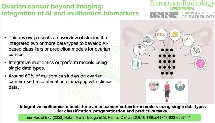
Ovarian cancer, often referred to as the “silent killer”, tends to show inconspicuous symptoms in the early stages, making timely diagnosis difficult. Detecting ovarian cancer at an early stage significantly increases the chances of effective treatment and therefore the chances of survival. This review study has shown that AI-based tools that rely on the integration of multi-omics data perform better

Read More →

Quo vadis Radiomics? Bibliometric analysis of 10-year Radiomics journey

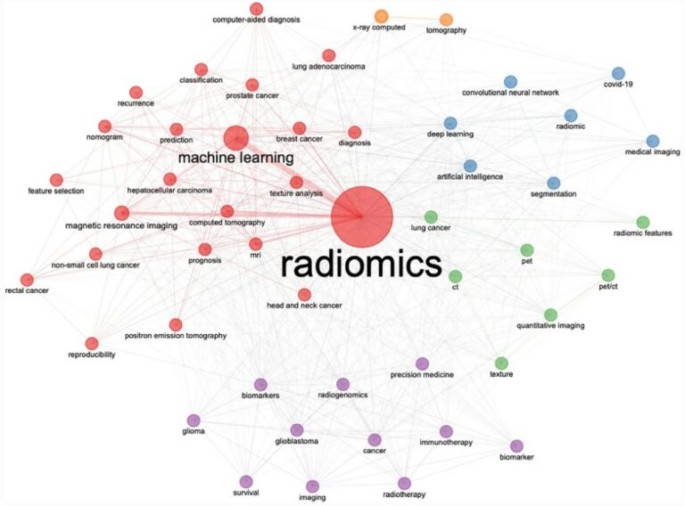
This study conducted a bibliometric analysis of radiomics ten years after the first work became available in March 2012. Throughout the analysis, the authors identified over 5,500 articles from almost 17,000 authors from over 900 different sources, highlighting developments within radiomics, its real-world applications, and tangible and intangible benefits. Key points ML-based bibliometric analysis is fundamental to detect unknown pattern

Read More →

Quality assurance for automatically generated contours with additional deep learning

This study explores the importance of quality assurance when deploying an automatic segmentation model. The authors of this study built a deep-learning model with the goal of estimating the quality of automatically generated contours. They found that the trained model can be used alongside automatic segmentation tools, thus ensuring quality and allowing intervention to prevent undesired segmentation behavior. Key points

Read More →

EuSoMII Radiomics Auditing Group Initiative: Review of RQS applications

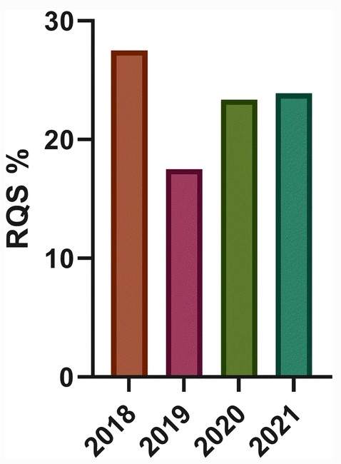
When the Radiomics Quality Score (RQS) was presented to the scientific community back in 2017, its authors aimed to introduce a tool for a rapid and effective evaluation of radiomics studies’ scientific/clinical merit. Conceived as a quality seal to be published alongside presented results and newly proposed radiomics models, the RQS has instead mostly been adopted by researchers as a

Read More →

Applications of machine learning for imaging-driven diagnosis of musculoskeletal malignancies

Due to the rare nature of musculoskeletal malignancies and the lack of imaging data associated with this cancer, the authors of this study aimed to investigate whether machine learning (ML) is already having an impact on imaging-driven diagnosis of musculoskeletal malignancies. The authors concluded that because of the limited amounts of data and no established large-scale networks between multiple national

Read More →

Can AI predict breast tumour response?

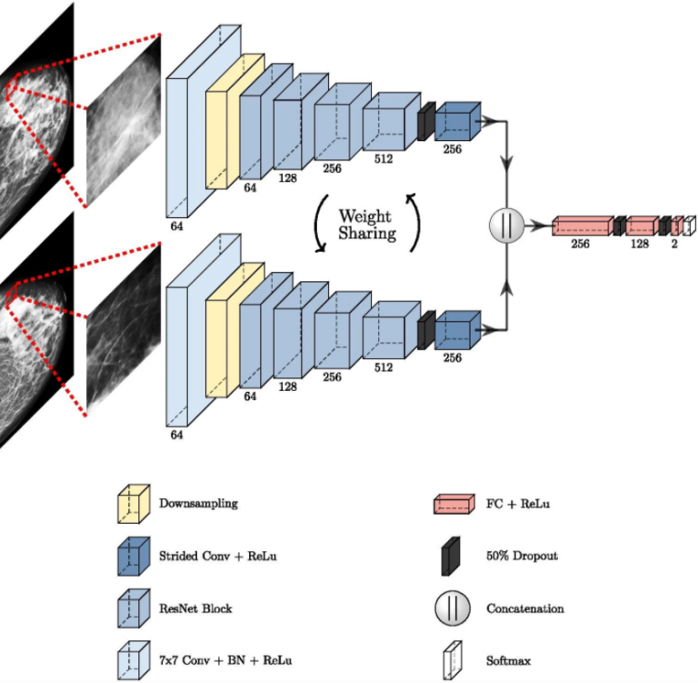
This proof of concept study examines using a deep learning-based method for the automatic analysis of digital mammograms as a tool to aid in the assessment of neoadjuvant chemotherapy (NACT) treatment response to breast cancer. The authors found that the initial AI performance was able to indicate the potential to aid in clinical decision-making, but in order to continue exploring

Read More →

An international survey on AI in radiology in 1041 radiologists and radiology residents part 2: expectations, hurdles to implementation, and education

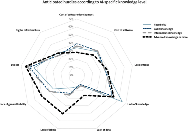
As a follow-up to part one of this international survey on artificial intelligence (AI), which surveyed over 1,000 radiologists and radiology residents and explored early adoption of AI, as well as radiologists’ perspectives on and fear of being replaced by these technologies, part 2 discusses the expectations of AI and the hurdles associated with implementation and education. Regardless of the

Read More →

Bridging the chasm: Education may turn Early Majority into Early Adopters

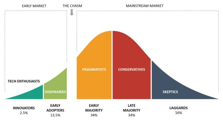
A while ago we came up with the idea to investigate the intersection between change management and artificial intelligence (AI) in radiology. We wanted to assess independent predictors for being a so-called ‘early adopter’, inspired by Roger’s Innovation Diffusion theory. An early adopter, coming right after the innovator, is someone who wants to take a risk without getting an immediate

Read More →

How can we combat multicenter variability in MR radiomics? Validation of a correction procedure

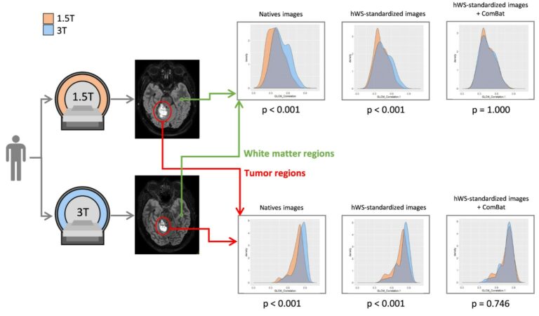
In this study, the authors extended the ComBat approach to provide a harmonization procedure that is applicable to any radiomic feature. They achieve this by combining image standardization with ComBat realignment, thus demonstrating that this could efficiently remove the scanner/protocol effect while preserving the individual variations in phantom, brain, and prostate MR scans. Key points Radiomic feature values obtained using

Read More →

Ovarian cancer beyond imaging: integration of AI and multiomics biomarkers

Ovarian cancer, often referred to as the “silent killer”, tends to show inconspicuous symptoms in the early stages, making timely diagnosis difficult. Detecting ovarian cancer at an early stage significantly increases the chances of effective treatment and therefore the chances of survival. This review study has shown that AI-based tools that rely on the integration of multi-omics data perform better

Read More →

Quo vadis Radiomics? Bibliometric analysis of 10-year Radiomics journey

This study conducted a bibliometric analysis of radiomics ten years after the first work became available in March 2012. Throughout the analysis, the authors identified over 5,500 articles from almost 17,000 authors from over 900 different sources, highlighting developments within radiomics, its real-world applications, and tangible and intangible benefits. Key points ML-based bibliometric analysis is fundamental to detect unknown pattern

Read More →

Quality assurance for automatically generated contours with additional deep learning

This study explores the importance of quality assurance when deploying an automatic segmentation model. The authors of this study built a deep-learning model with the goal of estimating the quality of automatically generated contours. They found that the trained model can be used alongside automatic segmentation tools, thus ensuring quality and allowing intervention to prevent undesired segmentation behavior. Key points

Read More →

EuSoMII Radiomics Auditing Group Initiative: Review of RQS applications

When the Radiomics Quality Score (RQS) was presented to the scientific community back in 2017, its authors aimed to introduce a tool for a rapid and effective evaluation of radiomics studies’ scientific/clinical merit. Conceived as a quality seal to be published alongside presented results and newly proposed radiomics models, the RQS has instead mostly been adopted by researchers as a

Read More →

Applications of machine learning for imaging-driven diagnosis of musculoskeletal malignancies

Due to the rare nature of musculoskeletal malignancies and the lack of imaging data associated with this cancer, the authors of this study aimed to investigate whether machine learning (ML) is already having an impact on imaging-driven diagnosis of musculoskeletal malignancies. The authors concluded that because of the limited amounts of data and no established large-scale networks between multiple national

Read More →

Can AI predict breast tumour response?

This proof of concept study examines using a deep learning-based method for the automatic analysis of digital mammograms as a tool to aid in the assessment of neoadjuvant chemotherapy (NACT) treatment response to breast cancer. The authors found that the initial AI performance was able to indicate the potential to aid in clinical decision-making, but in order to continue exploring

Read More →

An international survey on AI in radiology in 1041 radiologists and radiology residents part 2: expectations, hurdles to implementation, and education

As a follow-up to part one of this international survey on artificial intelligence (AI), which surveyed over 1,000 radiologists and radiology residents and explored early adoption of AI, as well as radiologists’ perspectives on and fear of being replaced by these technologies, part 2 discusses the expectations of AI and the hurdles associated with implementation and education. Regardless of the

Read More →

Bridging the chasm: Education may turn Early Majority into Early Adopters

A while ago we came up with the idea to investigate the intersection between change management and artificial intelligence (AI) in radiology. We wanted to assess independent predictors for being a so-called ‘early adopter’, inspired by Roger’s Innovation Diffusion theory. An early adopter, coming right after the innovator, is someone who wants to take a risk without getting an immediate

Read More →

How can we combat multicenter variability in MR radiomics? Validation of a correction procedure

In this study, the authors extended the ComBat approach to provide a harmonization procedure that is applicable to any radiomic feature. They achieve this by combining image standardization with ComBat realignment, thus demonstrating that this could efficiently remove the scanner/protocol effect while preserving the individual variations in phantom, brain, and prostate MR scans. Key points Radiomic feature values obtained using

Read More →

Become A Member Today!

You will have access to a wide range of benefits that can help you advance your career and stay up-to-date with the latest developments in the field of radiology. These benefits include access to educational resources, networking opportunities with other professionals in the field, opportunities to participate in research projects and clinical trials, and access to the latest technologies and techniques. 

Check out our different membership options.

If you don’t find a fitting membership send us an email here.

Membership

for radiologists, radiology residents, professionals of allied sciences (including radiographers/radiological technologists, nuclear medicine physicians, medical physicists, and data scientists) & professionals of allied sciences in training residing within the boundaries of Europe

  • Reduced registration fees for ECR 1
  • Reduced fees for the European School of Radiology (ESOR) 2
  • Option to participate in the European Diploma. 3
  • Free electronic access to the journal European Radiology 
  • Content e-mails for all ESR journals4
  • Updates on offers & events through our newsletters
  • Exclusive access to the ESR feed in Juisci

€ 11 /year

Yes! That is less than €1 per month.

Free membership

for radiologists, radiology residents or professionals of allied sciences engaged in practice, teaching or research residing outside Europe as well as individual qualified professionals with an interest in radiology and medical imaging who do not fulfil individual or all requirements for any other ESR membership category & former full members who have retired from all clinical practice
  • Reduced registration fees for ECR 1
  • Option to participate in the European Diploma. 3
  • Free electronic access to the journal European Radiology
  • Content e-mails for all ESR journals 4
  • Updates on offers & events through our newsletters
  • Exclusive access to the ESR feed in Juisci

€ 0

The best things in life are free.

ESR Friends

For students, company representatives or hospital managers etc.

  • Content e-mails for all 3 ESR journals 4
  • Updates on offers & events through our newsletters

€ 0

Friendship doesn’t cost a thing.

The membership type best fitting for you will be selected automatically during the application process.

Footnotes:

01

Reduced registration fees for ECR 2025:
Provided that ESR 2024 membership is activated and approved by August 31, 2024.

02
Not all activities included
03
Examination based on the ESR European Training Curriculum (radiologists or radiology residents).
04
European Radiology, Insights into Imaging, European Radiology Experimental.