MDCT

Welcome to the blog on Artificial Intelligence of
the European Society of Radiology

This blog aims at bringing educational and critical perspectives on AI to readers. It should help imaging professionals to learn and keep up to date with the technologies being developed in this rapidly evolving field.

Most used hashtags:

Latest posts

Deep learning image reconstruction to improve accuracy of iodine quantification and image quality in abdomen dual-energy CT

The aim of this study was to investigate the effect of deep learning image reconstruction (DLIR) on the accuracy of iodine quantification and image quality of dual-energy CT (DECT) compared to other reconstruction algorithms. The authors showed that the DLIR algorithm reduced image noise and variability of iodine concentration values when compared with other reconstruction algorithms. Key points In the

Read More →

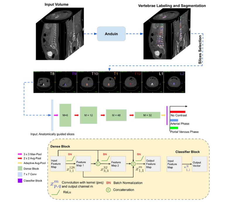
Artificial neural network detects contrast phase in MDCT

Automated extraction of novel biomarkers from routine imaging examinations, e.g., opportunistic CT, is a promising opportunity to extend current screening possibilities and to better guide individualized therapies. In theory, opportunistic CT can be used to obtain quantitative biomarker data from any tissue included in a routine examination. Over the past decade, AI has been used to develop many different automated

Read More →

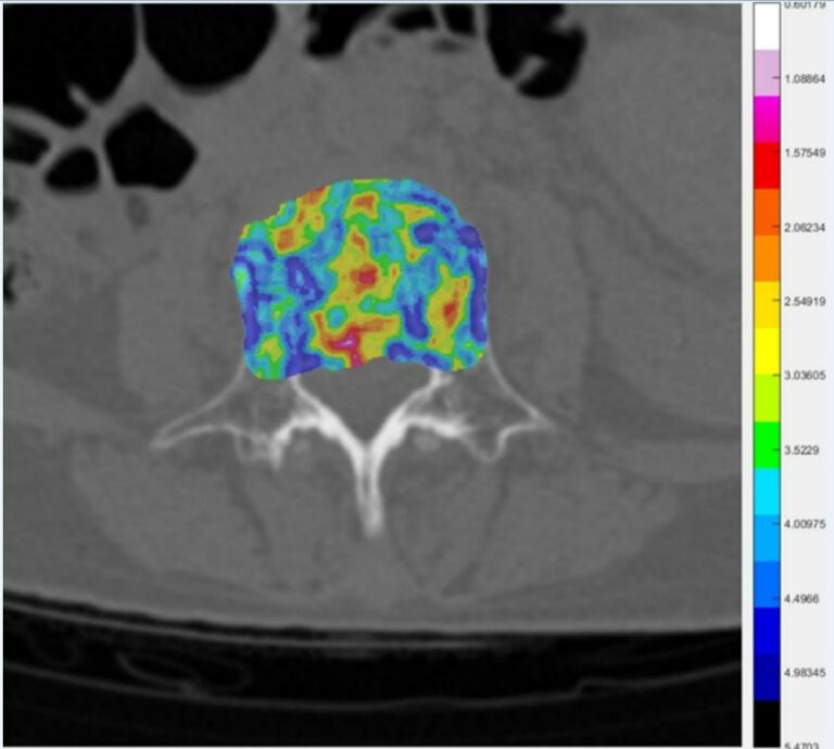
Combined radiomics-clinical model to predict malignancy of vertebral compression fractures on CT

The authors of this study aimed to develop and validate a combined radiomics-clinical model to predict malignancy of cerebral compression fractures on CT. This study comprised 165 patients with vertebral compression fractures who were allocated to training and validation cohorts. The authors were able to determine that the combined radiomics-clinical model integrating clinical parameters with radiomics score could predict malignancy

Read More →

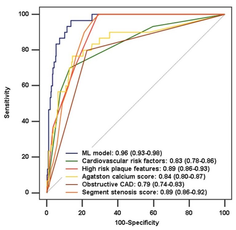
Improved long-term prognostic value of coronary CT angiography-derived plaque measures and clinical parameters on adverse cardiac outcome using machine learning

In recent years, artificial intelligence (AI) and, in particular, the application of machine learning (ML) algorithms, have become a new cornerstone in cardiovascular imaging with improved decision pathways, risk stratification, and outcome prediction in a more objective, reproducible, and rational manner. The integration of ML in daily routine clinical practice may hold potential to improve imaging workflow and to promote

Read More →

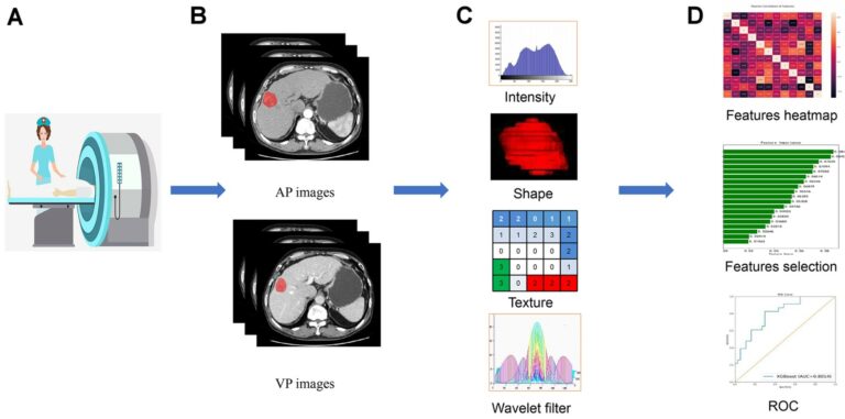
Preoperative prediction for pathological grade of hepatocellular carcinoma via machine learning–based radiomics

The purpose of this single-center retrospective study was to investigate the effectiveness of contrast-enhanced computed tomography (CECT)-based radiomic signatures for the preoperative prediction of pathological grades of hepatocellular carcinoma (HCC) via machine learning. The authors found that the radiomics signatures could non-invasively explore the underlying association between CECT images and pathological grades of HCC. Key points The radiomics signatures may

Read More →

Prediction of pulmonary pressure after Glenn shunts by computed tomography–based machine learning models

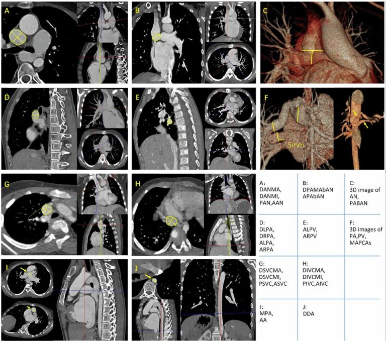
The aim of this retrospective study was to develop non-invasive machine learning (ML) classifiers for predicting post-Glenn shunt patients with low and high risks of a mean pulmonary arterial pressure above 15 mmHg, which was based on pre-operative cardiac CT. The study included 96 patients who underwent a bidirectional Glenn procedure. Key points Twenty-three candidate descriptors were manually extracted from

Read More →

Evaluation of an AI-based, automatic coronary artery calcium scoring software

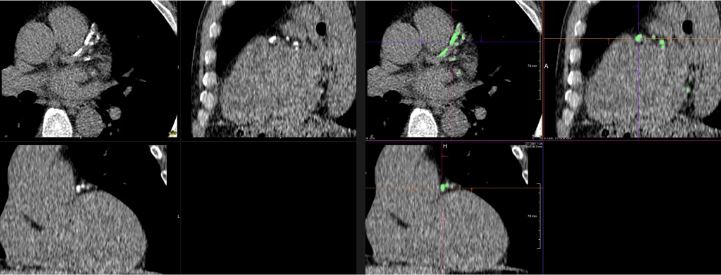
In this study, using a semi-automatic software as a reference, the authors aimed to evaluate an artificial intelligence (AI)-based, automatic coronary artery calcium (CAC) scoring software. This observational study included 315 non-contrast-enhanced calcium scoring computed tomography (CSCT) scans. The authors determined that there was a strong correlation between the automatic software and the semi-automatic software for three CAC scores and

Read More →

Noncontrast computer tomography–based radiomics model for predicting intracerebral hemorrhage expansion: preliminary findings and comparison with conventional radiological model

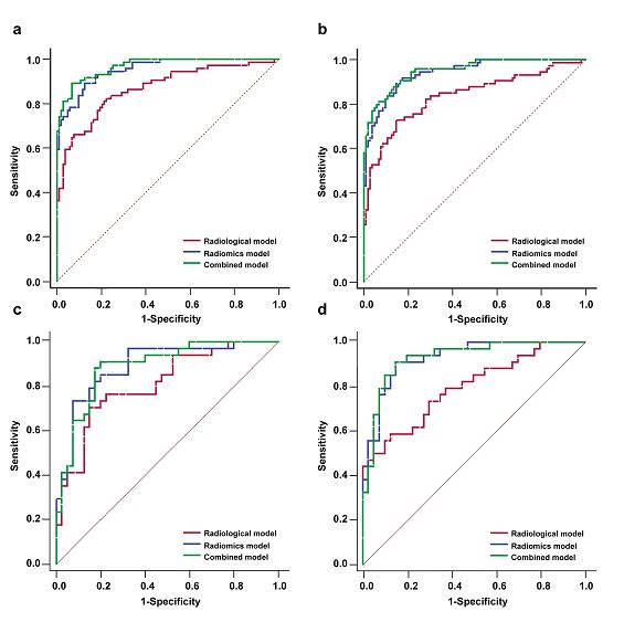
The authors developed a radiomics model for predicting hematoma expansion in patients with intracerebral haemorrhage (ICH) and compared its predictive performance with a conventional radiological feature-based model. Through retrospective analysis and noncontrast computed tomography (NCCT) assessment, it was found that an NCCT-based radiomics model showed better performance in the prediction of early hematoma expansion in ICH patients. Key points Radiomics

Read More →

Radiomics analysis using contrast-enhanced CT for preoperative prediction of occult peritoneal metastasis in advanced gastric cancer

The authors of this study constructed two multivariate logistic regression models and compared the diagnostic performance between the two of them via receiver operating characteristic analysis, discovering that CT radiomics analysis can provide valuable information for predicting occult peritoneal metastases in advanced gastric cancer. Key points Venous CT radiomics analysis provided valuable information for predicting occult peritoneal metastases in advanced

Read More →

Deep learning image reconstruction to improve accuracy of iodine quantification and image quality in abdomen dual-energy CT

The aim of this study was to investigate the effect of deep learning image reconstruction (DLIR) on the accuracy of iodine quantification and image quality of dual-energy CT (DECT) compared to other reconstruction algorithms. The authors showed that the DLIR algorithm reduced image noise and variability of iodine concentration values when compared with other reconstruction algorithms. Key points In the

Read More →

Artificial neural network detects contrast phase in MDCT

Automated extraction of novel biomarkers from routine imaging examinations, e.g., opportunistic CT, is a promising opportunity to extend current screening possibilities and to better guide individualized therapies. In theory, opportunistic CT can be used to obtain quantitative biomarker data from any tissue included in a routine examination. Over the past decade, AI has been used to develop many different automated

Read More →

Combined radiomics-clinical model to predict malignancy of vertebral compression fractures on CT

The authors of this study aimed to develop and validate a combined radiomics-clinical model to predict malignancy of cerebral compression fractures on CT. This study comprised 165 patients with vertebral compression fractures who were allocated to training and validation cohorts. The authors were able to determine that the combined radiomics-clinical model integrating clinical parameters with radiomics score could predict malignancy

Read More →

Improved long-term prognostic value of coronary CT angiography-derived plaque measures and clinical parameters on adverse cardiac outcome using machine learning

In recent years, artificial intelligence (AI) and, in particular, the application of machine learning (ML) algorithms, have become a new cornerstone in cardiovascular imaging with improved decision pathways, risk stratification, and outcome prediction in a more objective, reproducible, and rational manner. The integration of ML in daily routine clinical practice may hold potential to improve imaging workflow and to promote

Read More →

Preoperative prediction for pathological grade of hepatocellular carcinoma via machine learning–based radiomics

The purpose of this single-center retrospective study was to investigate the effectiveness of contrast-enhanced computed tomography (CECT)-based radiomic signatures for the preoperative prediction of pathological grades of hepatocellular carcinoma (HCC) via machine learning. The authors found that the radiomics signatures could non-invasively explore the underlying association between CECT images and pathological grades of HCC. Key points The radiomics signatures may

Read More →

Prediction of pulmonary pressure after Glenn shunts by computed tomography–based machine learning models

The aim of this retrospective study was to develop non-invasive machine learning (ML) classifiers for predicting post-Glenn shunt patients with low and high risks of a mean pulmonary arterial pressure above 15 mmHg, which was based on pre-operative cardiac CT. The study included 96 patients who underwent a bidirectional Glenn procedure. Key points Twenty-three candidate descriptors were manually extracted from

Read More →

Evaluation of an AI-based, automatic coronary artery calcium scoring software

In this study, using a semi-automatic software as a reference, the authors aimed to evaluate an artificial intelligence (AI)-based, automatic coronary artery calcium (CAC) scoring software. This observational study included 315 non-contrast-enhanced calcium scoring computed tomography (CSCT) scans. The authors determined that there was a strong correlation between the automatic software and the semi-automatic software for three CAC scores and

Read More →

Noncontrast computer tomography–based radiomics model for predicting intracerebral hemorrhage expansion: preliminary findings and comparison with conventional radiological model

The authors developed a radiomics model for predicting hematoma expansion in patients with intracerebral haemorrhage (ICH) and compared its predictive performance with a conventional radiological feature-based model. Through retrospective analysis and noncontrast computed tomography (NCCT) assessment, it was found that an NCCT-based radiomics model showed better performance in the prediction of early hematoma expansion in ICH patients. Key points Radiomics

Read More →

Radiomics analysis using contrast-enhanced CT for preoperative prediction of occult peritoneal metastasis in advanced gastric cancer

The authors of this study constructed two multivariate logistic regression models and compared the diagnostic performance between the two of them via receiver operating characteristic analysis, discovering that CT radiomics analysis can provide valuable information for predicting occult peritoneal metastases in advanced gastric cancer. Key points Venous CT radiomics analysis provided valuable information for predicting occult peritoneal metastases in advanced

Read More →

Become A Member Today!

You will have access to a wide range of benefits that can help you advance your career and stay up-to-date with the latest developments in the field of radiology. These benefits include access to educational resources, networking opportunities with other professionals in the field, opportunities to participate in research projects and clinical trials, and access to the latest technologies and techniques. 

Check out our different membership options.

If you don’t find a fitting membership send us an email here.

Membership

for radiologists, radiology residents, professionals of allied sciences (including radiographers/radiological technologists, nuclear medicine physicians, medical physicists, and data scientists) & professionals of allied sciences in training residing within the boundaries of Europe

  • Reduced registration fees for ECR 1
  • Reduced fees for the European School of Radiology (ESOR) 2
  • Option to participate in the European Diploma. 3
  • Free electronic access to the journal European Radiology 
  • Content e-mails for all ESR journals4
  • Updates on offers & events through our newsletters
  • Exclusive access to the ESR feed in Juisci

€ 11 /year

Yes! That is less than €1 per month.

Free membership

for radiologists, radiology residents or professionals of allied sciences engaged in practice, teaching or research residing outside Europe as well as individual qualified professionals with an interest in radiology and medical imaging who do not fulfil individual or all requirements for any other ESR membership category & former full members who have retired from all clinical practice
  • Reduced registration fees for ECR 1
  • Option to participate in the European Diploma. 3
  • Free electronic access to the journal European Radiology
  • Content e-mails for all ESR journals 4
  • Updates on offers & events through our newsletters
  • Exclusive access to the ESR feed in Juisci

€ 0

The best things in life are free.

ESR Friends

For students, company representatives or hospital managers etc.

  • Content e-mails for all 3 ESR journals 4
  • Updates on offers & events through our newsletters

€ 0

Friendship doesn’t cost a thing.

The membership type best fitting for you will be selected automatically during the application process.

Footnotes:

01

Reduced registration fees for ECR 2025:
Provided that ESR 2024 membership is activated and approved by August 31, 2024.

02
Not all activities included
03
Examination based on the ESR European Training Curriculum (radiologists or radiology residents).
04
European Radiology, Insights into Imaging, European Radiology Experimental.