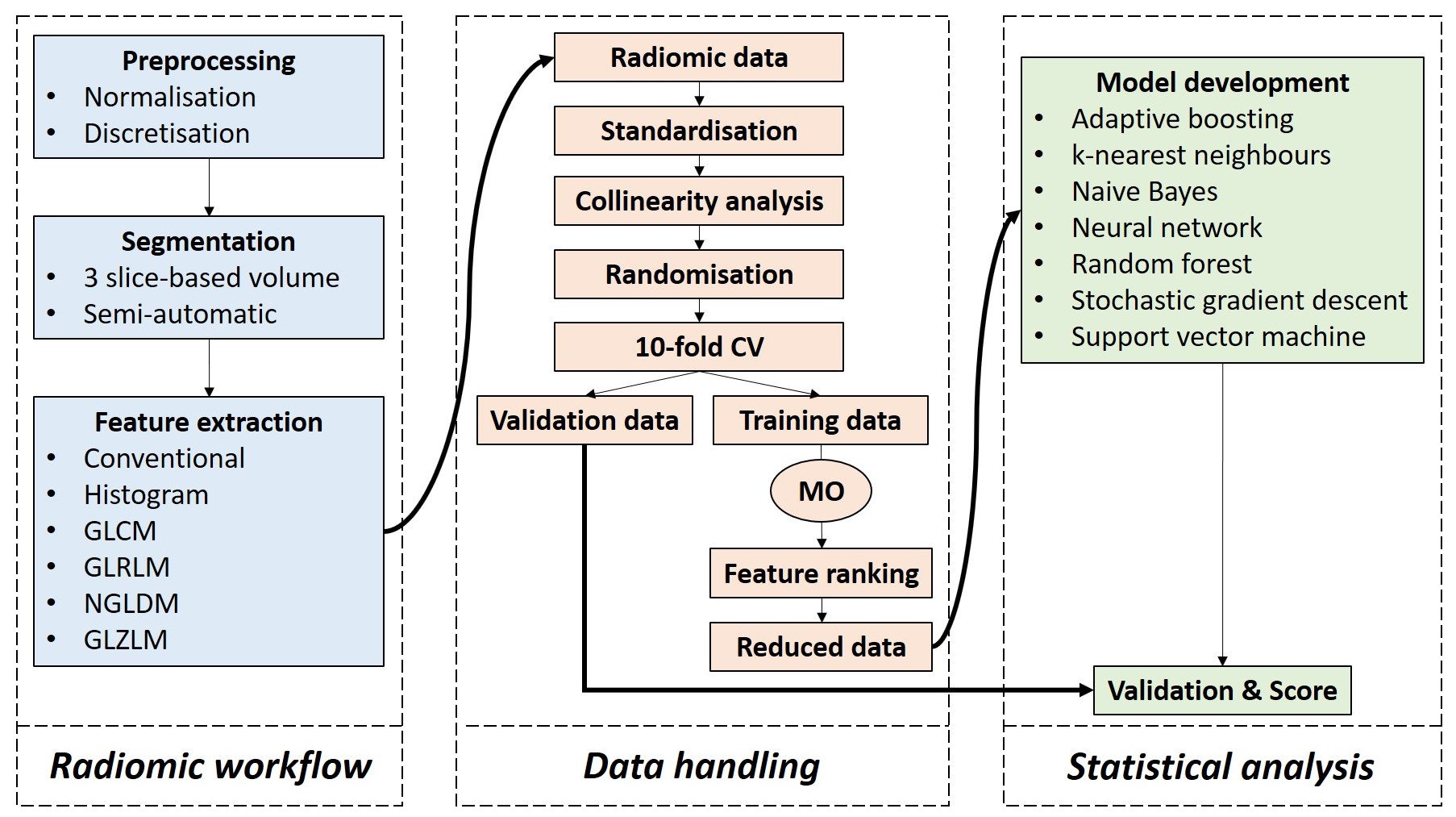
In this work, we aimed to evaluate the potential value of the machine learning (ML)–based MRI texture analysis for predicting 1p/19q codeletion status of lower-grade gliomas (LGG). This retrospective study was totally based on public data. We reduced the high-dimensionality of the radiomic data with collinearity analysis and ReliefF algorithm. Then, we used seven ML classifiers for the development of the models. Although there was no statistically significant difference among most of the classifiers, the artificial neural network had the highest rank.
Key points
- More than four-fifths of the lower-grade gliomas can be correctly classified with machine learning-based MRI texture analysis. Satisfying classification outcomes are not limited to a single algorithm.
- A few-slice-based volumetric segmentation technique would be a valid approach, providing satisfactory predictive textural information and avoiding excessive segmentation duration in clinical practice.
- Feature selection is sensitive to different patient data set samples so that each sampling leads to the selection of different feature subsets, which needs to be considered in future works.
Authors: Burak Kocak, Emine Sebnem Durmaz, Ece Ates, Ipek Sel, Saime Turgut Gunes, Ozlem Korkmaz Kaya, Amalya Zeynalova & Ozgur Kilickesmez


