biomarkers

Welcome to the blog on Artificial Intelligence of
the European Society of Radiology

This blog aims at bringing educational and critical perspectives on AI to readers. It should help imaging professionals to learn and keep up to date with the technologies being developed in this rapidly evolving field.

Most used hashtags:

Latest posts

Ovarian cancer beyond imaging: integration of AI and multiomics biomarkers

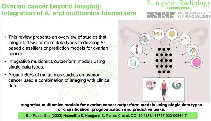
Ovarian cancer, often referred to as the “silent killer”, tends to show inconspicuous symptoms in the early stages, making timely diagnosis difficult. Detecting ovarian cancer at an early stage significantly increases the chances of effective treatment and therefore the chances of survival. This review study has shown that AI-based tools that rely on the integration of multi-omics data perform better

Read More →

QuantImage v2: physician-centered cloud platform for radiomics and machine learning research

The authors of this study proposed a physician-centered vision of radiomics research to help aid the translation of radiomics prediction models into clinical practice and workflow. Free-to-access radiomics tools and frameworks were reviewed to help identify best practices and gaps in the existing software. The authors designed QuantImage V2 (QI2) to help implement this vision and address any issues they

Read More →

Unlocking the Potential of Radiomics: Ongoing Challenges Are Revolving Around Methodology and Reproducibility

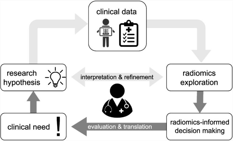
Over a decade in the making, the novel concept of radiomics has been silently brewing, promising to reshape the landscape of personalised and precision medicine. So, why has radiomics not made its clinical debut yet, despite its innovative and logical approach? The answer lies in the intricate world of advanced computation that forms the basis of radiomics, which in essence

Read More →

Using a multi-decoder water-fat separation neural network for liver PDFF estimation

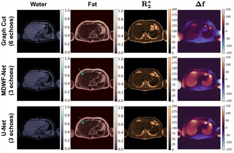
The authors of this study proposed a multi-task U-Net-based architecture to jointly estimate water-only and fat-only images. This approach allowed for the improvement in the estimation of water-fat images, enabling a reduction of the necessary echoes to achieve an accurate proton density fat fraction (PDFF) quantification. The proposed method was shown to be a reliable liver fat quantification tool for

Read More →

Can radiomics signatures predict tumor reponse of patients treated with chemotherapy and targeted therapy?

The authors of this retrospective study had the goal of evaluating the effectiveness of radiomics signatures in order to predict the tumor response of non-small cell lung cancer (NSCLC) patients who were treated with first-line chemotherapy, targeted therapy, or a combination of the two. The authors determined that radiomics signatures based on pre-treatment CT scans can accurately predict tumor response

Read More →

Radiomics in predicting treatment response in non-small-cell lung cancer: current status, challenges and future perspectives

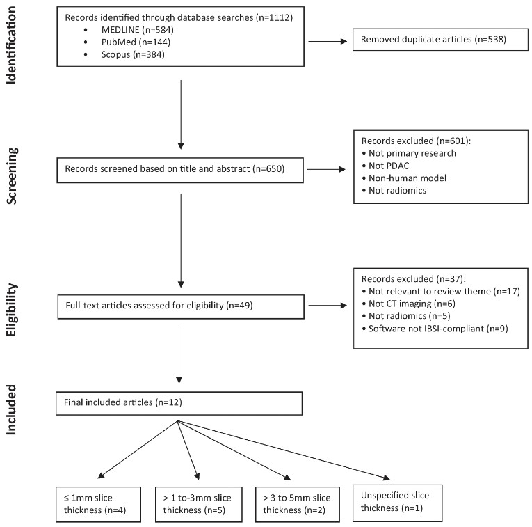
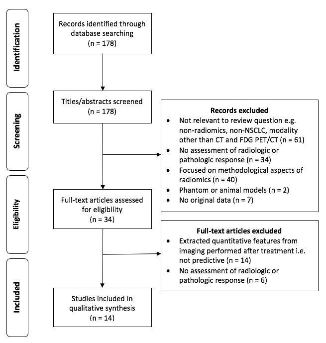
This literature review summarizes the current status and evaluates the scientific reporting quality of radiomics research in the prediction of treatment response in non-small-cell lung cancer (NSCLC). The authors performed this literature review through a comprehensive literature search using the PubMed database, screening a total of 178 articles for eligibility. Key points The included studies reported several promising radiomic markers

Read More →

Preoperative prediction for pathological grade of hepatocellular carcinoma via machine learning–based radiomics

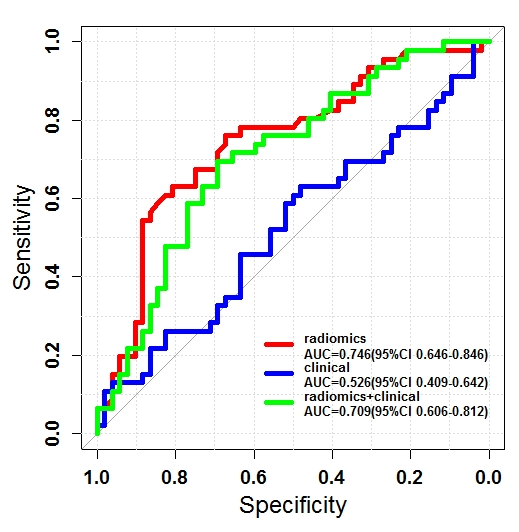
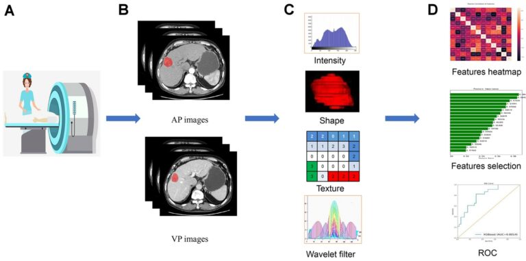
The purpose of this single-center retrospective study was to investigate the effectiveness of contrast-enhanced computed tomography (CECT)-based radiomic signatures for the preoperative prediction of pathological grades of hepatocellular carcinoma (HCC) via machine learning. The authors found that the radiomics signatures could non-invasively explore the underlying association between CECT images and pathological grades of HCC. Key points The radiomics signatures may

Read More →

Radiomics: the facts and the challenges of image analysis

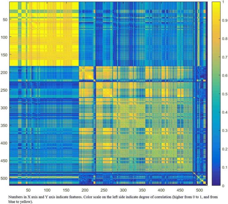
Radiomics is a complex multi-step process that can be considered as part of the more complex world of Artifical Intelligence (AI). The aim of radiomics is aiding clinical decision-making and outcome prediction for more personalized medicine. Each step of the radiomics process brings challenges that have to be considered; for example, segmentation is challenging because of reproducibility issues. Indeed, there

Read More →

Ovarian cancer beyond imaging: integration of AI and multiomics biomarkers

Ovarian cancer, often referred to as the “silent killer”, tends to show inconspicuous symptoms in the early stages, making timely diagnosis difficult. Detecting ovarian cancer at an early stage significantly increases the chances of effective treatment and therefore the chances of survival. This review study has shown that AI-based tools that rely on the integration of multi-omics data perform better

Read More →

QuantImage v2: physician-centered cloud platform for radiomics and machine learning research

The authors of this study proposed a physician-centered vision of radiomics research to help aid the translation of radiomics prediction models into clinical practice and workflow. Free-to-access radiomics tools and frameworks were reviewed to help identify best practices and gaps in the existing software. The authors designed QuantImage V2 (QI2) to help implement this vision and address any issues they

Read More →

Unlocking the Potential of Radiomics: Ongoing Challenges Are Revolving Around Methodology and Reproducibility

Over a decade in the making, the novel concept of radiomics has been silently brewing, promising to reshape the landscape of personalised and precision medicine. So, why has radiomics not made its clinical debut yet, despite its innovative and logical approach? The answer lies in the intricate world of advanced computation that forms the basis of radiomics, which in essence

Read More →

Using a multi-decoder water-fat separation neural network for liver PDFF estimation

The authors of this study proposed a multi-task U-Net-based architecture to jointly estimate water-only and fat-only images. This approach allowed for the improvement in the estimation of water-fat images, enabling a reduction of the necessary echoes to achieve an accurate proton density fat fraction (PDFF) quantification. The proposed method was shown to be a reliable liver fat quantification tool for

Read More →

Can radiomics signatures predict tumor reponse of patients treated with chemotherapy and targeted therapy?

The authors of this retrospective study had the goal of evaluating the effectiveness of radiomics signatures in order to predict the tumor response of non-small cell lung cancer (NSCLC) patients who were treated with first-line chemotherapy, targeted therapy, or a combination of the two. The authors determined that radiomics signatures based on pre-treatment CT scans can accurately predict tumor response

Read More →

Radiomics in predicting treatment response in non-small-cell lung cancer: current status, challenges and future perspectives

This literature review summarizes the current status and evaluates the scientific reporting quality of radiomics research in the prediction of treatment response in non-small-cell lung cancer (NSCLC). The authors performed this literature review through a comprehensive literature search using the PubMed database, screening a total of 178 articles for eligibility. Key points The included studies reported several promising radiomic markers

Read More →

Preoperative prediction for pathological grade of hepatocellular carcinoma via machine learning–based radiomics

The purpose of this single-center retrospective study was to investigate the effectiveness of contrast-enhanced computed tomography (CECT)-based radiomic signatures for the preoperative prediction of pathological grades of hepatocellular carcinoma (HCC) via machine learning. The authors found that the radiomics signatures could non-invasively explore the underlying association between CECT images and pathological grades of HCC. Key points The radiomics signatures may

Read More →

Radiomics: the facts and the challenges of image analysis

Radiomics is a complex multi-step process that can be considered as part of the more complex world of Artifical Intelligence (AI). The aim of radiomics is aiding clinical decision-making and outcome prediction for more personalized medicine. Each step of the radiomics process brings challenges that have to be considered; for example, segmentation is challenging because of reproducibility issues. Indeed, there

Read More →

Become A Member Today!

You will have access to a wide range of benefits that can help you advance your career and stay up-to-date with the latest developments in the field of radiology. These benefits include access to educational resources, networking opportunities with other professionals in the field, opportunities to participate in research projects and clinical trials, and access to the latest technologies and techniques. 

Check out our different membership options.

If you don’t find a fitting membership send us an email here.

Membership

for radiologists, radiology residents, professionals of allied sciences (including radiographers/radiological technologists, nuclear medicine physicians, medical physicists, and data scientists) & professionals of allied sciences in training residing within the boundaries of Europe

  • Reduced registration fees for ECR 1
  • Reduced fees for the European School of Radiology (ESOR) 2
  • Option to participate in the European Diploma. 3
  • Free electronic access to the journal European Radiology 
  • Content e-mails for all ESR journals4
  • Updates on offers & events through our newsletters
  • Exclusive access to the ESR feed in Juisci

€ 11 /year

Yes! That is less than €1 per month.

Free membership

for radiologists, radiology residents or professionals of allied sciences engaged in practice, teaching or research residing outside Europe as well as individual qualified professionals with an interest in radiology and medical imaging who do not fulfil individual or all requirements for any other ESR membership category & former full members who have retired from all clinical practice
  • Reduced registration fees for ECR 1
  • Option to participate in the European Diploma. 3
  • Free electronic access to the journal European Radiology
  • Content e-mails for all ESR journals 4
  • Updates on offers & events through our newsletters
  • Exclusive access to the ESR feed in Juisci

€ 0

The best things in life are free.

ESR Friends

For students, company representatives or hospital managers etc.

  • Content e-mails for all 3 ESR journals 4
  • Updates on offers & events through our newsletters

€ 0

Friendship doesn’t cost a thing.

The membership type best fitting for you will be selected automatically during the application process.

Footnotes:

01

Reduced registration fees for ECR 2025:
Provided that ESR 2024 membership is activated and approved by August 31, 2024.

02
Not all activities included
03
Examination based on the ESR European Training Curriculum (radiologists or radiology residents).
04
European Radiology, Insights into Imaging, European Radiology Experimental.