reproducibility of results

Welcome to the blog on Artificial Intelligence of
the European Society of Radiology

This blog aims at bringing educational and critical perspectives on AI to readers. It should help imaging professionals to learn and keep up to date with the technologies being developed in this rapidly evolving field.

Most used hashtags:

Latest posts

Sharing is Caring – Promoting Radiomics Research Transparency and Sustainability

Radiomics, a rapidly growing and innovative field in medical imaging, extracts detailed features from medical scans that could play a pivotal future adjunct role in patient care. However, the clinical adoption of radiomics is stalled by significant research heterogeneity and reproducibility issues [1]. The complex radiomics pipeline, requiring multidisciplinary expertise, often lacks transparency regarding sharing crucial details like software tools

Read More →

Reproducibility of radiomics quality score: an intra- and inter-rater reliability study

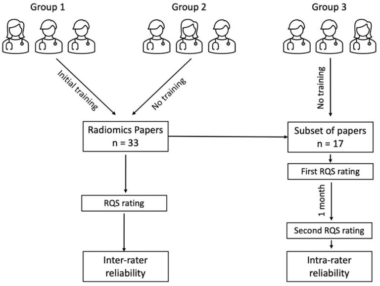
The rapidly evolving field of radiomics research holds the potential to revolutionize medicine by transforming diagnostic images into quantifiable data. Assessing the quality of radiomics research is challenging and requires reliable tools for standardization. Our results, published in European Radiology, have revealed some room for improvement regarding the reproducibility of the widely used Radiomics Quality Score (RQS). We recruited multiple

Read More →

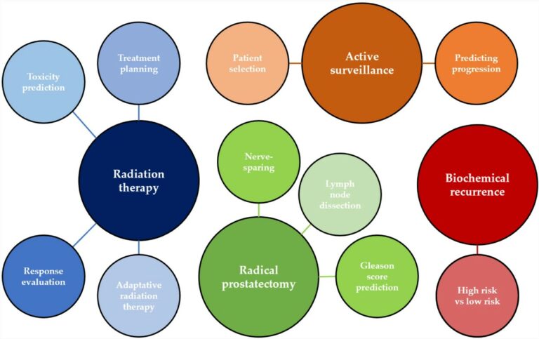
Beyond diagnosis: is there a role for radiomics in prostate cancer management?

At present, therapeutic and prognostic recommendations for prostate cancer (PCa) predominantly hinge on risk-stratification tools that are built upon clinical parameters. Recent evidence indicates that incorporating imaging can enhance the precision of prognostic models based on clinical factors. However, challenges like subjective interpretation, variability in image analysis, and the absence of reliable quantitative measures need to be overcome to fully

Read More →

Unlocking the Potential of Radiomics: Ongoing Challenges Are Revolving Around Methodology and Reproducibility

Over a decade in the making, the novel concept of radiomics has been silently brewing, promising to reshape the landscape of personalised and precision medicine. So, why has radiomics not made its clinical debut yet, despite its innovative and logical approach? The answer lies in the intricate world of advanced computation that forms the basis of radiomics, which in essence

Read More →

Deep learning image reconstruction improves DECT image quality

The purpose of this phantom study was the compare the image quality of a deep learning image reconstruction (DLIR) algorithm and conventional iterative reconstruction (IR) algorithms in dual-energy CT (DECT) as well as assess the impact that these algorithms have on radiomics robustness. The authors determined that the new DLIR algorithm does in fact improve the quality of DECT images

Read More →

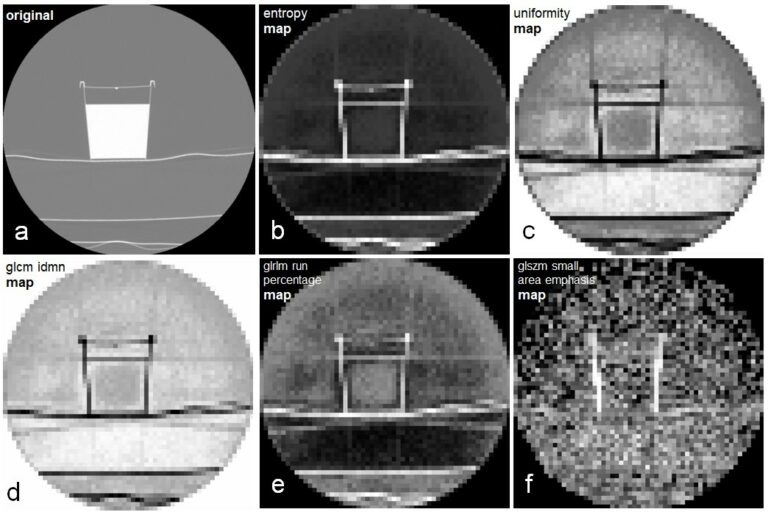
Using parametric feature maps to enhance the stability of CT radiomics

It is known that the reproducibility of radiomic features is influenced by myriad factors, one of which is the size of the segmented volume. We hypothesized that parametric maps calculated with a fixed voxel size could address this issue. To test the hypothesis, we conducted a phantom study and could show that the stability across different volumes of interest sizes

Read More →

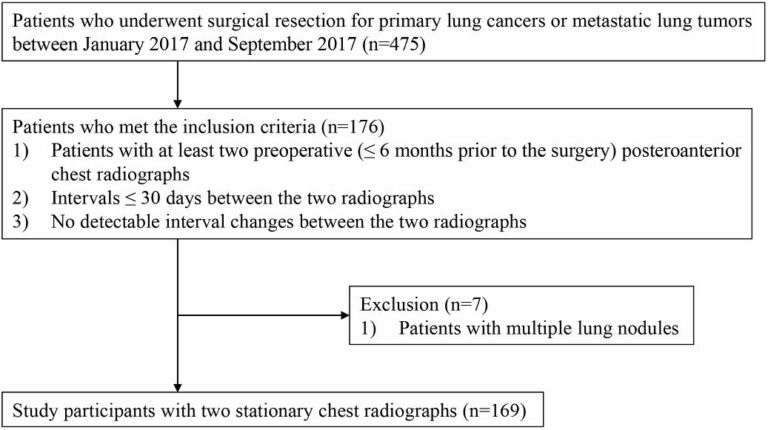
Test-retest reproducibility of a deep learning–based automatic detection algorithm for the chest radiograph

The authors of this retrospective study performed test-retest reproducibility analyses for a deep learning-based automatic detection algorithm (DLAD) using two stationary chest radiographs with short-term intervals, in order to analyze influential factors on test-retest variations. The test, which included patients with pulmonary nodules resected in 2017, showed that DLAD was robust to the test-retest variation. Key points The deep learning–based

Read More →

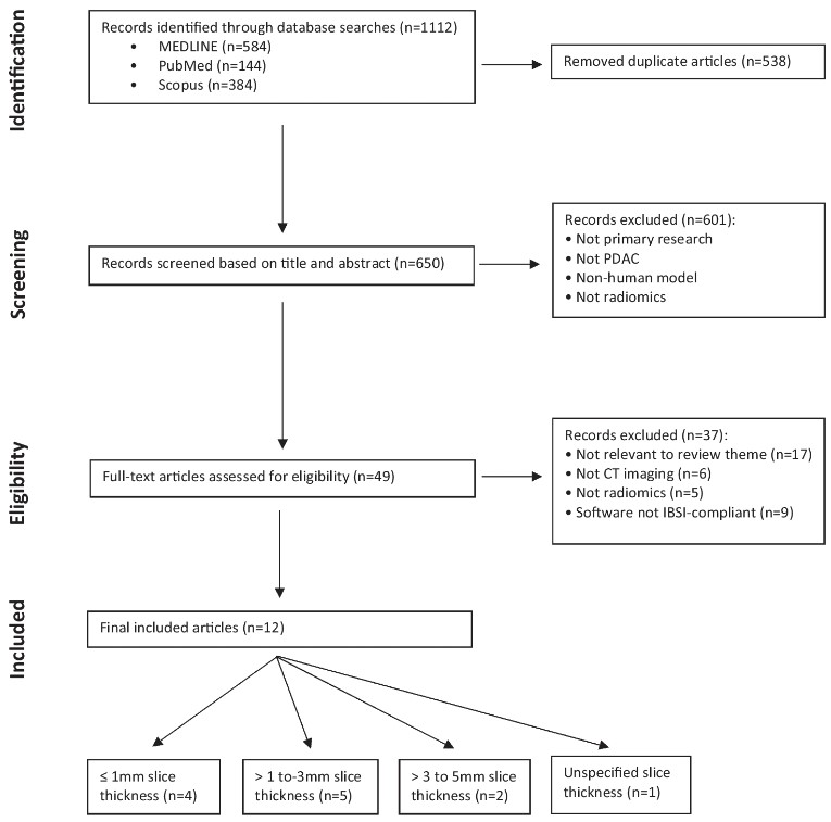
Radiomic feature reproducibility in contrast-enhanced CT of the pancreas is affected by variabilities in scan parameters and manual segmentation

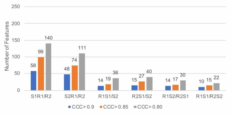
The goal of this study was to measure the reproducibility of radiomic features in pancreatic parenchyma and ductal adenocarcinomas in patients who underwent consecutive contrast-enhanced computed tomography (CECT) scans. A majority of features were not reproducible (as defined by a concordance correlation coefficient of greater than 0.9) when comparing their values across consecutive CECTs obtained within a two week period.

Read More →

Sharing is Caring – Promoting Radiomics Research Transparency and Sustainability

Radiomics, a rapidly growing and innovative field in medical imaging, extracts detailed features from medical scans that could play a pivotal future adjunct role in patient care. However, the clinical adoption of radiomics is stalled by significant research heterogeneity and reproducibility issues [1]. The complex radiomics pipeline, requiring multidisciplinary expertise, often lacks transparency regarding sharing crucial details like software tools

Read More →

Reproducibility of radiomics quality score: an intra- and inter-rater reliability study

The rapidly evolving field of radiomics research holds the potential to revolutionize medicine by transforming diagnostic images into quantifiable data. Assessing the quality of radiomics research is challenging and requires reliable tools for standardization. Our results, published in European Radiology, have revealed some room for improvement regarding the reproducibility of the widely used Radiomics Quality Score (RQS). We recruited multiple

Read More →

Beyond diagnosis: is there a role for radiomics in prostate cancer management?

At present, therapeutic and prognostic recommendations for prostate cancer (PCa) predominantly hinge on risk-stratification tools that are built upon clinical parameters. Recent evidence indicates that incorporating imaging can enhance the precision of prognostic models based on clinical factors. However, challenges like subjective interpretation, variability in image analysis, and the absence of reliable quantitative measures need to be overcome to fully

Read More →

Unlocking the Potential of Radiomics: Ongoing Challenges Are Revolving Around Methodology and Reproducibility

Over a decade in the making, the novel concept of radiomics has been silently brewing, promising to reshape the landscape of personalised and precision medicine. So, why has radiomics not made its clinical debut yet, despite its innovative and logical approach? The answer lies in the intricate world of advanced computation that forms the basis of radiomics, which in essence

Read More →

Deep learning image reconstruction improves DECT image quality

The purpose of this phantom study was the compare the image quality of a deep learning image reconstruction (DLIR) algorithm and conventional iterative reconstruction (IR) algorithms in dual-energy CT (DECT) as well as assess the impact that these algorithms have on radiomics robustness. The authors determined that the new DLIR algorithm does in fact improve the quality of DECT images

Read More →

Using parametric feature maps to enhance the stability of CT radiomics

It is known that the reproducibility of radiomic features is influenced by myriad factors, one of which is the size of the segmented volume. We hypothesized that parametric maps calculated with a fixed voxel size could address this issue. To test the hypothesis, we conducted a phantom study and could show that the stability across different volumes of interest sizes

Read More →

Test-retest reproducibility of a deep learning–based automatic detection algorithm for the chest radiograph

The authors of this retrospective study performed test-retest reproducibility analyses for a deep learning-based automatic detection algorithm (DLAD) using two stationary chest radiographs with short-term intervals, in order to analyze influential factors on test-retest variations. The test, which included patients with pulmonary nodules resected in 2017, showed that DLAD was robust to the test-retest variation. Key points The deep learning–based

Read More →

Radiomic feature reproducibility in contrast-enhanced CT of the pancreas is affected by variabilities in scan parameters and manual segmentation

The goal of this study was to measure the reproducibility of radiomic features in pancreatic parenchyma and ductal adenocarcinomas in patients who underwent consecutive contrast-enhanced computed tomography (CECT) scans. A majority of features were not reproducible (as defined by a concordance correlation coefficient of greater than 0.9) when comparing their values across consecutive CECTs obtained within a two week period.

Read More →

Become A Member Today!

You will have access to a wide range of benefits that can help you advance your career and stay up-to-date with the latest developments in the field of radiology. These benefits include access to educational resources, networking opportunities with other professionals in the field, opportunities to participate in research projects and clinical trials, and access to the latest technologies and techniques. 

Check out our different membership options.

If you don’t find a fitting membership send us an email here.

Membership

for radiologists, radiology residents, professionals of allied sciences (including radiographers/radiological technologists, nuclear medicine physicians, medical physicists, and data scientists) & professionals of allied sciences in training residing within the boundaries of Europe

  • Reduced registration fees for ECR 1
  • Reduced fees for the European School of Radiology (ESOR) 2
  • Option to participate in the European Diploma. 3
  • Free electronic access to the journal European Radiology 
  • Content e-mails for all ESR journals4
  • Updates on offers & events through our newsletters
  • Exclusive access to the ESR feed in Juisci

€ 11 /year

Yes! That is less than €1 per month.

Free membership

for radiologists, radiology residents or professionals of allied sciences engaged in practice, teaching or research residing outside Europe as well as individual qualified professionals with an interest in radiology and medical imaging who do not fulfil individual or all requirements for any other ESR membership category & former full members who have retired from all clinical practice
  • Reduced registration fees for ECR 1
  • Option to participate in the European Diploma. 3
  • Free electronic access to the journal European Radiology
  • Content e-mails for all ESR journals 4
  • Updates on offers & events through our newsletters
  • Exclusive access to the ESR feed in Juisci

€ 0

The best things in life are free.

ESR Friends

For students, company representatives or hospital managers etc.

  • Content e-mails for all 3 ESR journals 4
  • Updates on offers & events through our newsletters

€ 0

Friendship doesn’t cost a thing.

The membership type best fitting for you will be selected automatically during the application process.

Footnotes:

01

Reduced registration fees for ECR 2025:
Provided that ESR 2024 membership is activated and approved by August 31, 2024.

Reduced registration fees for ECR 2026:
Provided that ESR 2025 membership is activated and approved by August 31, 2025.

02
Not all activities included
03
Examination based on the ESR European Training Curriculum (radiologists or radiology residents).
04
European Radiology, Insights into Imaging, European Radiology Experimental.