AI Blog

Welcome to the blog on Artificial Intelligence of
the European Society of Radiology

This blog aims at bringing educational and critical perspectives on AI to readers. It should help imaging professionals to learn and keep up to date with the technologies being developed in this rapidly evolving field.

Most used hashtags:

Latest posts

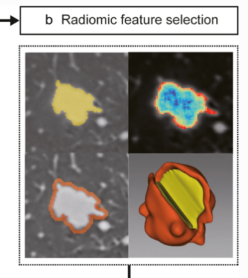
Radiomics in the prediction of disease-free survival in early-stage squamous cervical cancer

The authors of this study conducted multiparametric magnetic resonance imaging (MRI)-derived radiomics based on multi-scale tumor region in order to predict disease-free survival (DFS) in a cohort of 191 patients with early-stage squamous cervical cancer (ESSCC). They were able to conclude that multiparametric MRI-derived radiomics based on multi-scale tumor region can in fact aid in the prediction of DFS for

Read More →

Creating a training set for AI from initial segmentations of airways

An important challenge in the use of artificial intelligence (AI) for medical image segmentation tasks is the lack of high-quality, scan protocol-specific datasets. AI performs best on narrow tasks with homogenous specifications. Thus, pre-trained models may be inadequate for use in centre-specific studies if the scan protocols do not match. For the airway segmentation task in the Imaging in Lifelines

Read More →

A deep learning algorithm for VHD diagnosis and evaluation

This study aims to develop and validate a deep learning-based automatic chest radiograph (CXR) cardiovascular border (CB) analysis algorithm (CB_auto) in order to diagnose and quantitatively evaluate valvular heart disease (VHD). The authors found that the CB_auto system, in coordination with the deep learning algorithm, provided highly reliable CB measurements, which, in turn, can be useful, not long daily clinical

Read More →

Deep learning radiomics in prediction of NAC response

For breast cancer, the standard of treatment for most patients is neoadjuvant chemotherapy (NAC), but response rates may vary among patients, causing delays in appropriate treatment. The authors of this prospective study aimed to investigate the feasibility of deep learning radiomics (DLR) in the prediction of NAC response at an early stage of breast cancer treatment. The authors found that

Read More →

Misunderstandings on equitable and inequitable biases in machine learning

In this reply to a ‘Letter to the editor‘, the authors seek to correct a few misunderstandings relating to equitable and inequitable biases in machine learning and radiology that were mentioned by the authors of the letter, in which topics such as cultural biases and ‘socially related’ cognitive biases were discussed, as well as how to deal with these biases.

Read More →

Deep learning assesses additional radiation dose in overscanning

Following the COVID-19 pandemic, the number of chest CT examinations has dramatically increased, which will undeniably impact public medical exposure. Overscanning, i.e., scanning unnecessary regions in the axial field-of-view, causes noticeable excessive radiation dose to patients undergoing chest CT examinations. The manual procedure of selecting the scan range based on anterior-posterior or lateral localizers is prone to human error in

Read More →

Can AI predict breast tumour response?

This proof of concept study examines using a deep learning-based method for the automatic analysis of digital mammograms as a tool to aid in the assessment of neoadjuvant chemotherapy (NACT) treatment response to breast cancer. The authors found that the initial AI performance was able to indicate the potential to aid in clinical decision-making, but in order to continue exploring

Read More →

Radiomic nomogram predicts pathology invasiveness

The purpose of this retrospective diagnostic study was to develop and validate a preoperative CT-based nomogram combined with radiomic and clinical-radiological signatures to distinguish preinvasive lesions from pulmonary invasive lesions. The authors were able to demonstrate that a nomogram constructed by identified clinical-radiological signatures and combined radiomic signatures has the potential to precisely predict pathology invasiveness. Key points The radiomic

Read More →

Automated AI to predict survival post cystectomy

In this study, the authors developed a fully automated artificial intelligence (AI)-based image analysis tool for segmenting skeletal muscle of the torso and calculating the muscle volume. The authors were able to determine that the fully automated AI-based image analysis software was able to segment the skeletal muscle volume in over 97% of patients who were planning to undergo radical

Read More →

Integrated AI model aids ultrasonographers

In this study, the authors aimed to develop an explainable ultrasound (US) computer-assisted diagnostic (CAD) model for suspicious thyroid nodules by retrospectively analyzing over 2,900 solid or almost-solid thyroid nodules. A deep learning model and a multiple risk features learning ensemble model were then used to train the US images of 2,794 thyroid nodules. An integrated AI model was generated

Read More →

Become A Member Today!

You will have access to a wide range of benefits that can help you advance your career and stay up-to-date with the latest developments in the field of radiology. These benefits include access to educational resources, networking opportunities with other professionals in the field, opportunities to participate in research projects and clinical trials, and access to the latest technologies and techniques. 

Check out our different membership options.

If you don’t find a fitting membership send us an email here.

Membership

for radiologists, radiology residents, professionals of allied sciences (including radiographers/radiological technologists, nuclear medicine physicians, medical physicists, and data scientists) & professionals of allied sciences in training residing within the boundaries of Europe

  • Reduced registration fees for ECR 1
  • Reduced fees for the European School of Radiology (ESOR) 2
  • Exclusive option to participate in the European Diploma. 3
  • Free electronic access to the journal European Radiology 4
  • Content e-mails for all ESR journals
  • Updates on offers & events through our newsletters
  • Exclusive access to the ESR feed in Juisci

€ 11 /year

Yes! That is less than €1 per month.

Free membership

for radiologists, radiology residents or professionals of allied sciences engaged in practice, teaching or research residing outside Europe as well as individual qualified professionals with an interest in radiology and medical imaging who do not fulfil individual or all requirements for any other ESR membership category & former full members who have retired from all clinical practice
  • Reduced registration fees for ECR 1
  • Free electronic access to the journal European Radiology
  • Content e-mails for all 3 ESR journals 4
  • Updates on offers & events through our newsletters
  • Exclusive access to the ESR feed in Juisci

€ 0

The best things in life are free.

ESR Friends

For students, company representatives or hospital managers etc.

  • Content e-mails for all 3 ESR journals 4
  • Updates on offers & events through our newsletters

€ 0

Friendship doesn’t cost a thing.

The membership type best fitting for you will be selected automatically during the application process.

Footnotes:

01

Reduced registration fees for ECR 2024:
Provided that ESR 2023 membership is activated and approved by August 31, 2023.

Reduced registration fees for ECR 2025:
Provided that ESR 2024 membership is activated and approved by August 31, 2024.

02
Not all activities included
03
Examination based on the ESR European Training Curriculum (radiologists or radiology residents).
04
European Radiology, Insights into Imaging, European Radiology Experimental.