AI Blog

Welcome to the blog on Artificial Intelligence of
the European Society of Radiology

This blog aims at bringing educational and critical perspectives on AI to readers. It should help imaging professionals to learn and keep up to date with the technologies being developed in this rapidly evolving field.

Most used hashtags:

Latest posts

Using a multiple classifier system for lesion detection and classification in breast MRI analysis

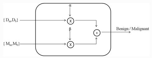
In breast magnetic resonance imaging (MRI) analysis for lesion detection and classification, radiologists agree that both morphological and dynamic features are important to differentiate benign from malignant lesions. This study proposes a multiple classifier system (MCS) to classify breast lesions on dynamic contrast-enhanced MRI (DCE-MRI) combining morphological features and dynamic information. The data gained through testing showed that an MCS

Read More →

Prediction of rupture risk in anterior communicating artery aneurysms

This article attempts to predict the rupture risk in anterior communicating artery (ACOM) aneurysms by using a two-layer feed-forward artificial neural network (ANN). To improve ANN efficiency, an adaptive synthetic (ADASYN) sampling approach was applied to generate more synthetic data for unruptured aneurysms. Based on the results, the conclusion is that this ANN presents good performance and offers a valuable

Read More →

The Rise of Augmented Radiology as an enabler to achieve Precision Health

The field of medical imaging has witnessed a revolution thanks to the digital transformation, innovation and availability of advanced clinical applications. New imaging techniques are helping radiologists, oncologists, and other diagnosticians with greater anatomical and clinical details, highlighting the need for fast access to imaging reports and results, evidence-based collaborative peer review and collaboration, and predictive intelligence. A pioneer in

Read More →

Convolutional neural networks: an overview and application in radiology

Numerous domains, including radiology, have shown interest in convolutional neural network (CNN) – a class of artificial neural networks that has become dominant in various computer vision tasks. It is designed to automatically and adaptively learn spatial hierarchies of features through backpropagation by using multiple building blocks, such as convolution layers, pooling layers, and fully connected layers. This review article

Read More →

Treat artificial intelligence as disruptive technology

Radiologists tend to be afraid of artificial intelligence (AI), but they should demystify it and take it for what it is, i.e. disruptive technology, according to Frank Lexa, a professor of radiology at the University of Arizona and an expert in medical leadership. “AI tools improve at the speed of light, and it’s unclear whether radiologists will keep on playing

Read More →

Big data, artificial intelligence, and structured reporting

Imagine you wanted to teach a baby to differentiate a ball from a dog. How would you do that? Intuitively it would make sense to repeatedly point at them while naming each object accordingly. But how about, e.g. if instead of saying “dog” or “ball”, you would only name the specific breed of dog without using the word “dog”? Maybe

Read More →

A survey of medical students’ attitude toward AI

The aim of this survey was to assess undergraduate medical students’ attitudes towards artificial intelligence (AI) in radiology and medicine. A web-based questionnaire consisting of various sections aiming to evaluate the students’ prior knowledge of AI in radiology and beyond was sent out to students at three major medical schools. The survey showed that undergraduate medical students do not worry

Read More →

AI for CT Image Reconstruction – A Great Opportunity

It seems like everybody in the radiology field is now working with Artificial Intelligence (AI) in one way or another. Especially here in the Silicon Valley, the number of ventures that develop AI algorithms is increasing by the day. What are these newly developed algorithms focusing on? Most of them try to solve a specific problem, and they do that

Read More →

Radiomics of liver MRI predict metastases in mice

This study aimed to investigate whether any texture features show a correlation with intrahepatic tumor growth before the metastasis is visible to the human eye. For the purposes of the study, eight mice were injected intraportally with syngeneic MC-38 colon cancer cells and two mice were injected with phosphate-buffered saline (sham controls). Magnetic resonance imaging (MRI) and texture analysis were

Read More →

Comparing computer- and human-extracted imaging phenotypes

This retrospective study sought to investigate if computer-extracted magnetic resonance imaging (MRI) phenotypes of breast cancer could replicate human-extracted size and Breast Imaging-Reporting and Data System (BI-RADS) imaging phenotypes using MRI data from The Cancer Genome Atlas (TCGA) project of the National Cancer Institute. Upon obtaining the results, it was possible to conclude that quantitative radiomics of breast cancer may replicate human-extracted tumour size

Read More →

Become A Member Today!

You will have access to a wide range of benefits that can help you advance your career and stay up-to-date with the latest developments in the field of radiology. These benefits include access to educational resources, networking opportunities with other professionals in the field, opportunities to participate in research projects and clinical trials, and access to the latest technologies and techniques. 

Check out our different membership options.

If you don’t find a fitting membership send us an email here.

Membership

for radiologists, radiology residents, professionals of allied sciences (including radiographers/radiological technologists, nuclear medicine physicians, medical physicists, and data scientists) & professionals of allied sciences in training residing within the boundaries of Europe

  • Reduced registration fees for ECR 1
  • Reduced fees for the European School of Radiology (ESOR) 2
  • Exclusive option to participate in the European Diploma. 3
  • Free electronic access to the journal European Radiology 4
  • Content e-mails for all ESR journals
  • Updates on offers & events through our newsletters
  • Exclusive access to the ESR feed in Juisci

€ 11 /year

Yes! That is less than €1 per month.

Free membership

for radiologists, radiology residents or professionals of allied sciences engaged in practice, teaching or research residing outside Europe as well as individual qualified professionals with an interest in radiology and medical imaging who do not fulfil individual or all requirements for any other ESR membership category & former full members who have retired from all clinical practice
  • Reduced registration fees for ECR 1
  • Free electronic access to the journal European Radiology
  • Content e-mails for all 3 ESR journals 4
  • Updates on offers & events through our newsletters
  • Exclusive access to the ESR feed in Juisci

€ 0

The best things in life are free.

ESR Friends

For students, company representatives or hospital managers etc.

  • Content e-mails for all 3 ESR journals 4
  • Updates on offers & events through our newsletters

€ 0

Friendship doesn’t cost a thing.

The membership type best fitting for you will be selected automatically during the application process.

Footnotes:

01

Reduced registration fees for ECR 2024:
Provided that ESR 2023 membership is activated and approved by August 31, 2023.

Reduced registration fees for ECR 2025:
Provided that ESR 2024 membership is activated and approved by August 31, 2024.

02
Not all activities included
03
Examination based on the ESR European Training Curriculum (radiologists or radiology residents).
04
European Radiology, Insights into Imaging, European Radiology Experimental.