mri

Welcome to the blog on Artificial Intelligence of
the European Society of Radiology

This blog aims at bringing educational and critical perspectives on AI to readers. It should help imaging professionals to learn and keep up to date with the technologies being developed in this rapidly evolving field.

Most used hashtags:

Latest posts

Machine learning classifiers vs. Experienced radiologists: Predicting Gleason pattern 4 prostate cancer

The authors of this study recognized the potential of multiparametric positron emission tomography/magnetic resonance imaging (mpPET/MRI) for detecting and classifying breast lesions. To better understand computer-aided segmentation and diagnosis (CAD) features, the authors introduced a data-driven machine learning approach for a CAD system that enables the assessment of the relevance of mpPET/MRI features on segmentation and classification accuracy. Key points:

Read More →

Automatic segmentation and classification of breast lesions through identification of informative multiparametric PET/MRI features

The authors of this study recognized the potential of multiparametric positron emission tomography/magnetic resonance imaging (mpPET/MRI) for detecting and classifying breast lesions. To better understand computer-aided segmentation and diagnosis (CAD) features, the authors introduced a data-driven machine learning approach for a CAD system that enables the assessment of the relevance of mpPET/MRI features on segmentation and classification accuracy. Key points:

Read More →

Using radiomics for the preoperative estimation of pathological grade in bladder cancer tumors

The goal of this study was to develop and authenticate an MRI-based radiomics strategy for the preoperative estimation of pathological grade in bladder cancer (BCa) tumors. The obtained radiomic features were evaluated for diagnostic abilities using receiver operating characteristic (ROC) curve analysis and compared using the DeLong test. The authors then performed validation using 30 consecutive patients and were able

Read More →

Is there a future for deep learning-based generative models in musculoskeletal radiology?

This article sought to investigate the potential of generative models in the field of MRI of the spine, and did so by performing clinically relevant benchmark cases. The interest in generative models, which are computer programs that are able to generate novel data, as opposed to classifying or processing existing data, is due to the fact that considerable technological innovations

Read More →

Using a multiple classifier system for lesion detection and classification in breast MRI analysis

In breast magnetic resonance imaging (MRI) analysis for lesion detection and classification, radiologists agree that both morphological and dynamic features are important to differentiate benign from malignant lesions. This study proposes a multiple classifier system (MCS) to classify breast lesions on dynamic contrast-enhanced MRI (DCE-MRI) combining morphological features and dynamic information. The data gained through testing showed that an MCS

Read More →

Radiomics of liver MRI predict metastases in mice

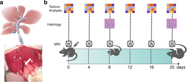
This study aimed to investigate whether any texture features show a correlation with intrahepatic tumor growth before the metastasis is visible to the human eye. For the purposes of the study, eight mice were injected intraportally with syngeneic MC-38 colon cancer cells and two mice were injected with phosphate-buffered saline (sham controls). Magnetic resonance imaging (MRI) and texture analysis were

Read More →

Comparing computer- and human-extracted imaging phenotypes

This retrospective study sought to investigate if computer-extracted magnetic resonance imaging (MRI) phenotypes of breast cancer could replicate human-extracted size and Breast Imaging-Reporting and Data System (BI-RADS) imaging phenotypes using MRI data from The Cancer Genome Atlas (TCGA) project of the National Cancer Institute. Upon obtaining the results, it was possible to conclude that quantitative radiomics of breast cancer may replicate human-extracted tumour size

Read More →

Machine learning classifiers vs. Experienced radiologists: Predicting Gleason pattern 4 prostate cancer

The authors of this study recognized the potential of multiparametric positron emission tomography/magnetic resonance imaging (mpPET/MRI) for detecting and classifying breast lesions. To better understand computer-aided segmentation and diagnosis (CAD) features, the authors introduced a data-driven machine learning approach for a CAD system that enables the assessment of the relevance of mpPET/MRI features on segmentation and classification accuracy. Key points:

Read More →

Automatic segmentation and classification of breast lesions through identification of informative multiparametric PET/MRI features

The authors of this study recognized the potential of multiparametric positron emission tomography/magnetic resonance imaging (mpPET/MRI) for detecting and classifying breast lesions. To better understand computer-aided segmentation and diagnosis (CAD) features, the authors introduced a data-driven machine learning approach for a CAD system that enables the assessment of the relevance of mpPET/MRI features on segmentation and classification accuracy. Key points:

Read More →

Using radiomics for the preoperative estimation of pathological grade in bladder cancer tumors

The goal of this study was to develop and authenticate an MRI-based radiomics strategy for the preoperative estimation of pathological grade in bladder cancer (BCa) tumors. The obtained radiomic features were evaluated for diagnostic abilities using receiver operating characteristic (ROC) curve analysis and compared using the DeLong test. The authors then performed validation using 30 consecutive patients and were able

Read More →

Is there a future for deep learning-based generative models in musculoskeletal radiology?

This article sought to investigate the potential of generative models in the field of MRI of the spine, and did so by performing clinically relevant benchmark cases. The interest in generative models, which are computer programs that are able to generate novel data, as opposed to classifying or processing existing data, is due to the fact that considerable technological innovations

Read More →

Using a multiple classifier system for lesion detection and classification in breast MRI analysis

In breast magnetic resonance imaging (MRI) analysis for lesion detection and classification, radiologists agree that both morphological and dynamic features are important to differentiate benign from malignant lesions. This study proposes a multiple classifier system (MCS) to classify breast lesions on dynamic contrast-enhanced MRI (DCE-MRI) combining morphological features and dynamic information. The data gained through testing showed that an MCS

Read More →

Radiomics of liver MRI predict metastases in mice

This study aimed to investigate whether any texture features show a correlation with intrahepatic tumor growth before the metastasis is visible to the human eye. For the purposes of the study, eight mice were injected intraportally with syngeneic MC-38 colon cancer cells and two mice were injected with phosphate-buffered saline (sham controls). Magnetic resonance imaging (MRI) and texture analysis were

Read More →

Comparing computer- and human-extracted imaging phenotypes

This retrospective study sought to investigate if computer-extracted magnetic resonance imaging (MRI) phenotypes of breast cancer could replicate human-extracted size and Breast Imaging-Reporting and Data System (BI-RADS) imaging phenotypes using MRI data from The Cancer Genome Atlas (TCGA) project of the National Cancer Institute. Upon obtaining the results, it was possible to conclude that quantitative radiomics of breast cancer may replicate human-extracted tumour size

Read More →

Become A Member Today!

You will have access to a wide range of benefits that can help you advance your career and stay up-to-date with the latest developments in the field of radiology. These benefits include access to educational resources, networking opportunities with other professionals in the field, opportunities to participate in research projects and clinical trials, and access to the latest technologies and techniques. 

Check out our different membership options.

If you don’t find a fitting membership send us an email here.

Membership

for radiologists, radiology residents, professionals of allied sciences (including radiographers/radiological technologists, nuclear medicine physicians, medical physicists, and data scientists) & professionals of allied sciences in training residing within the boundaries of Europe

  • Reduced registration fees for ECR 1
  • Reduced fees for the European School of Radiology (ESOR) 2
  • Exclusive option to participate in the European Diploma. 3
  • Free electronic access to the journal European Radiology 4
  • Content e-mails for all ESR journals
  • Updates on offers & events through our newsletters
  • Exclusive access to the ESR feed in Juisci

€ 11 /year

Yes! That is less than €1 per month.

Free membership

for radiologists, radiology residents or professionals of allied sciences engaged in practice, teaching or research residing outside Europe as well as individual qualified professionals with an interest in radiology and medical imaging who do not fulfil individual or all requirements for any other ESR membership category & former full members who have retired from all clinical practice
  • Reduced registration fees for ECR 1
  • Free electronic access to the journal European Radiology
  • Content e-mails for all 3 ESR journals 4
  • Updates on offers & events through our newsletters
  • Exclusive access to the ESR feed in Juisci

€ 0

The best things in life are free.

ESR Friends

For students, company representatives or hospital managers etc.

  • Content e-mails for all 3 ESR journals 4
  • Updates on offers & events through our newsletters

€ 0

Friendship doesn’t cost a thing.

The membership type best fitting for you will be selected automatically during the application process.

Footnotes:

01

Reduced registration fees for ECR 2024:
Provided that ESR 2023 membership is activated and approved by August 31, 2023.

Reduced registration fees for ECR 2025:
Provided that ESR 2024 membership is activated and approved by August 31, 2024.

02
Not all activities included
03
Examination based on the ESR European Training Curriculum (radiologists or radiology residents).
04
European Radiology, Insights into Imaging, European Radiology Experimental.