AI Blog

Welcome to the blog on Artificial Intelligence of
the European Society of Radiology

This blog aims at bringing educational and critical perspectives on AI to readers. It should help imaging professionals to learn and keep up to date with the technologies being developed in this rapidly evolving field.

Most used hashtags:

Latest posts

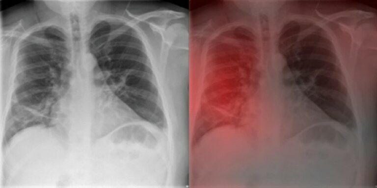
AI-aided software for detecting visible clinically significant prostate cancer on mpMRI

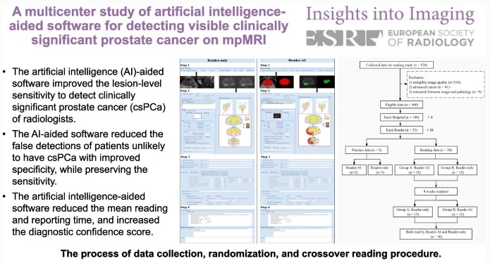
This study seeks to determine if artificial intelligence (AI)-based software can improve radiologists’ performance when detecting clinically significant prostate cancer. Sixteen radiologists from four hospitals participated and were assigned 30 cases, half without AI and half with AI. The authors determined that the AI software improves the performance of radiologists by reducing false positive detection of prostate cancer patients while

Read More →

Using a multi-decoder water-fat separation neural network for liver PDFF estimation

The authors of this study proposed a multi-task U-Net-based architecture to jointly estimate water-only and fat-only images. This approach allowed for the improvement in the estimation of water-fat images, enabling a reduction of the necessary echoes to achieve an accurate proton density fat fraction (PDFF) quantification. The proposed method was shown to be a reliable liver fat quantification tool for

Read More →

Hepatic steatosis increases liver volume

Our recent research published in European Radiology aimed to evaluate the impact of hepatic steatosis (HS) on liver volume by conducting a retrospective analysis of 1,038 living liver donors. We measured liver volume on gadoxetic acid-enhanced hepatobiliary phase MR images and proton density fat fraction (PDFF). Our results showed that HS leads to a 4.4% increase in liver volume per

Read More →

Radiologists’ performance improved by algorithmic transparency and interpretability measures

This study’s aim was to evaluate the perception of various types of artificial intelligence-based assistance and the interaction of radiologists with the algorithm’s predictions and certainty measures. As was consistent with previous research, the authors determined that human performance was superior to both groups when combined with AI. They also found an increase in trust in the algorithm’s performance when

Read More →

Machine learning and radiomics differentiate Crohn’s disease and ulcerative colitis

This retrospective study investigated whether volumetric visceral adipose tissue (VAT) features that were extracted using radiomics and three-dimensional convolutional neural network (3D-CNN) approaches are effective when differentiating Crohn’s disease (CD) and ulcerative colitis (UC). The authors concluded that VAT-based deep learning and radiomics features were able to achieve fair accuracy in differentiating CD from UC. Key points High-output feature data

Read More →

Covid-19 early detection: Neural Networks vs. Radiologists

The COVID-19 pandemic not only made an impact on the discipline of radiology as a whole but also on how we use specific tools in its detection. This was especially seen in the role of chest radiography when it was utilized as a diagnostic tool at the beginning of the pandemic “when microbiological resources were scarce,” evolving into its use

Read More →

Deep learning image reconstruction improves DECT image quality

The purpose of this phantom study was the compare the image quality of a deep learning image reconstruction (DLIR) algorithm and conventional iterative reconstruction (IR) algorithms in dual-energy CT (DECT) as well as assess the impact that these algorithms have on radiomics robustness. The authors determined that the new DLIR algorithm does in fact improve the quality of DECT images

Read More →

Automated vetting of radiology referrals: exploring NLP and ML approaches

As computed tomography (CT) sees an increase in utilization, inappropriate imaging has been seen as a significant concern; however, manual justification audits of radiology referrals are extremely time-consuming and carry a heavy financial burden. Therefore, the authors of this study aimed to retrospectively audit the justification of brain CT referrals by using natural language processing (NLP) and traditional machine learning

Read More →

T2-weighted MRI-based radiomics discriminati between benign and borderline epithelial ovarian tumors

The authors of this study investigated whether radiomics based on T2-weighted MRI was able to discriminate between benign and borderline epithelial ovarian tumors (EOTs) preoperatively. They were able to show that it can provide critical diagnostic information in the discrimination between benign and borderline EOTs, showing that there is potential to aid personalized treatment options. Key points T2-weighted MRI-based radiomics

Read More →

Super-resolution application of GAN on brain time-of-flight MR angiography

This study aimed to develop a generative adversarial network (GAN) model to improve the image resolution of brain time-of-flight MR angiography (TOF-MRA), as well as evaluate the image quality and diagnostic utility of the reconstructed images. The results showed that an optimized GAN could significantly improve the image quality and vessel visibility of low-resolution 3D TOF-MRA while maintaining equivalent sensitivity

Read More →

Become A Member Today!

You will have access to a wide range of benefits that can help you advance your career and stay up-to-date with the latest developments in the field of radiology. These benefits include access to educational resources, networking opportunities with other professionals in the field, opportunities to participate in research projects and clinical trials, and access to the latest technologies and techniques. 

Check out our different membership options.

If you don’t find a fitting membership send us an email here.

Membership

for radiologists, radiology residents, professionals of allied sciences (including radiographers/radiological technologists, nuclear medicine physicians, medical physicists, and data scientists) & professionals of allied sciences in training residing within the boundaries of Europe

  • Reduced registration fees for ECR 1
  • Reduced fees for the European School of Radiology (ESOR) 2
  • Exclusive option to participate in the European Diploma. 3
  • Free electronic access to the journal European Radiology 4
  • Content e-mails for all ESR journals
  • Updates on offers & events through our newsletters
  • Exclusive access to the ESR feed in Juisci

€ 11 /year

Yes! That is less than €1 per month.

Free membership

for radiologists, radiology residents or professionals of allied sciences engaged in practice, teaching or research residing outside Europe as well as individual qualified professionals with an interest in radiology and medical imaging who do not fulfil individual or all requirements for any other ESR membership category & former full members who have retired from all clinical practice
  • Reduced registration fees for ECR 1
  • Free electronic access to the journal European Radiology
  • Content e-mails for all 3 ESR journals 4
  • Updates on offers & events through our newsletters
  • Exclusive access to the ESR feed in Juisci

€ 0

The best things in life are free.

ESR Friends

For students, company representatives or hospital managers etc.

  • Content e-mails for all 3 ESR journals 4
  • Updates on offers & events through our newsletters

€ 0

Friendship doesn’t cost a thing.

The membership type best fitting for you will be selected automatically during the application process.

Footnotes:

01

Reduced registration fees for ECR 2024:
Provided that ESR 2023 membership is activated and approved by August 31, 2023.

Reduced registration fees for ECR 2025:
Provided that ESR 2024 membership is activated and approved by August 31, 2024.

02
Not all activities included
03
Examination based on the ESR European Training Curriculum (radiologists or radiology residents).
04
European Radiology, Insights into Imaging, European Radiology Experimental.