AI Blog

Welcome to the blog on Artificial Intelligence of
the European Society of Radiology

This blog aims at bringing educational and critical perspectives on AI to readers. It should help imaging professionals to learn and keep up to date with the technologies being developed in this rapidly evolving field.

Most used hashtags:

Latest posts

Prediction of lipomatous soft tissue malignancy on MRI

This study, which included a cohort of 145 patients affected by lipomatous soft tissue tumours, aimed to compare the performances of MRI radiomic machine learning analysis with that of deep learning in order to predict malignancy in patients with lipomas oratypical lipomatous tumours. The authors were able to show that batch-effect corrected machine learning and radiomics approaches outperformed deep learning-based

Read More →

Opportunistic screening for osteoporosis and osteopenia using machine learning

Dr. Rizwan Aslam, of the University of California, San Francisco (UCSF), presented an abstract at RSNA in 2008 which showed that it was possible to screen for osteoporosis from CT colonoscopy scans while I was a resident at UCSF. Subsequently, other papers showed that there were strong correlations between CT measurements and the measurements obtained from dual-energy x-ray absorptiometry (DXA).

Read More →

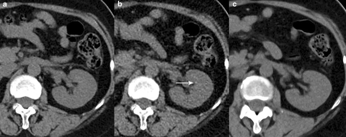
Application of deep learning reconstruction in the diagnosis of renal calculi

In this study, the authors evaluated the diagnostic capability, image quality, and radiation dose of abdominal ultra-low-dose CT (ULDCT) with deep learning reconstruction (DLR) in the detection of renal calculi, which are a common urological disease and normally detected by CT. They were able to find that ULDCT combined with DLR could significantly reduce radiation dose, decrease image noise, and

Read More →

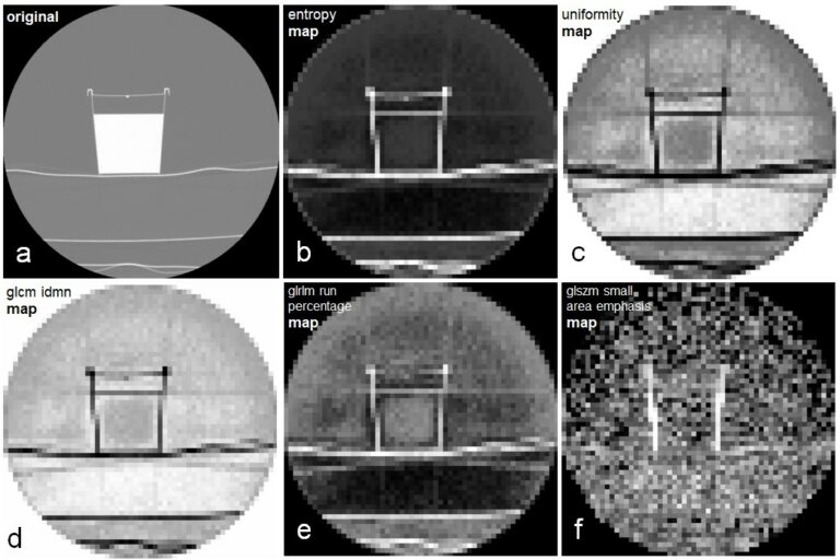
Using parametric feature maps to enhance the stability of CT radiomics

It is known that the reproducibility of radiomic features is influenced by myriad factors, one of which is the size of the segmented volume. We hypothesized that parametric maps calculated with a fixed voxel size could address this issue. To test the hypothesis, we conducted a phantom study and could show that the stability across different volumes of interest sizes

Read More →

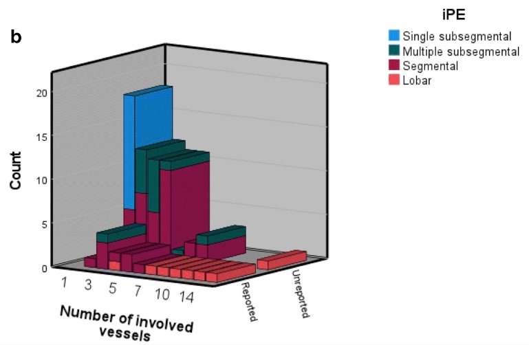
Cancer-associated incidental pulmonary embolism – underreporting and the potential role of AI

Pulmonary embolism (PE) is a common complication in patients with cancer. A significant number of all PE are diagnosed incidentally (incidental PE, iPE) in CT scans performed for staging or treatment response evaluation. Thousands of CT scans are performed for this indication every year in Halmstad, a regional hospital in Region Halland in southern Sweden, and we anticipate that in

Read More →

Deep learning image reconstruction to improve accuracy of iodine quantification and image quality in abdomen dual-energy CT

The aim of this study was to investigate the effect of deep learning image reconstruction (DLIR) on the accuracy of iodine quantification and image quality of dual-energy CT (DECT) compared to other reconstruction algorithms. The authors showed that the DLIR algorithm reduced image noise and variability of iodine concentration values when compared with other reconstruction algorithms. Key points In the

Read More →

Reproducibility of AI models in head CT

The authors of this study reviewed research on AI algorithms relating to computed tomography (CT) of the head in order to verify to what degree it is true that AI software for applications in radiology must be transferable to other real-world problems. It was discovered that current research on AI for head CT is rarely reproducible, does not match with

Read More →

The role of radiomics in the detection of lymph node metastases

The authors of this retrospective analysis looked at the role that radiomics played when applied to contrast-enhanced computed tomography (CT) in detecting lymph node (LN) metastases in lung cancer patients compared to 18F-fluorodeoxyglucose positron emission tomography (PET)/CT as a reference. The authors determined that radiomics showed good discrimination power, regardless of the modelling technique, in detecting LN metastases in lung

Read More →

Machine learning–based radiomics classifies parotid tumors using morphological MRI

This comparative study aimed to evaluate the effectiveness of machine learning models based on morphological MRI radiomics in the classification of parotid tumors. The authors developed three-step machine learning models with extreme gradient boosting (XGBoost), support vector machine (SVM), and decision tree (DT) algorithms in order to classify the parotid neoplasms into four subtypes. The study was able to demonstrate

Read More →

Communicating with patients in the age of online portals

Future generations of patients are very likely to get more involved in decisions regarding their healthcare according to the motto “nothing about me without me”. The deployment of electronic patient portals increasingly allows patients throughout Europe to consult and share their medical data, including radiology reports and images, securely and timely online. Technical solutions and rules for releasing reports and

Read More →

Become A Member Today!

You will have access to a wide range of benefits that can help you advance your career and stay up-to-date with the latest developments in the field of radiology. These benefits include access to educational resources, networking opportunities with other professionals in the field, opportunities to participate in research projects and clinical trials, and access to the latest technologies and techniques. 

Check out our different membership options.

If you don’t find a fitting membership send us an email here.

Membership

for radiologists, radiology residents, professionals of allied sciences (including radiographers/radiological technologists, nuclear medicine physicians, medical physicists, and data scientists) & professionals of allied sciences in training residing within the boundaries of Europe

  • Reduced registration fees for ECR 1
  • Reduced fees for the European School of Radiology (ESOR) 2
  • Exclusive option to participate in the European Diploma. 3
  • Free electronic access to the journal European Radiology 4
  • Content e-mails for all ESR journals
  • Updates on offers & events through our newsletters
  • Exclusive access to the ESR feed in Juisci

€ 11 /year

Yes! That is less than €1 per month.

Free membership

for radiologists, radiology residents or professionals of allied sciences engaged in practice, teaching or research residing outside Europe as well as individual qualified professionals with an interest in radiology and medical imaging who do not fulfil individual or all requirements for any other ESR membership category & former full members who have retired from all clinical practice
  • Reduced registration fees for ECR 1
  • Free electronic access to the journal European Radiology
  • Content e-mails for all 3 ESR journals 4
  • Updates on offers & events through our newsletters
  • Exclusive access to the ESR feed in Juisci

€ 0

The best things in life are free.

ESR Friends

For students, company representatives or hospital managers etc.

  • Content e-mails for all 3 ESR journals 4
  • Updates on offers & events through our newsletters

€ 0

Friendship doesn’t cost a thing.

The membership type best fitting for you will be selected automatically during the application process.

Footnotes:

01

Reduced registration fees for ECR 2024:
Provided that ESR 2023 membership is activated and approved by August 31, 2023.

Reduced registration fees for ECR 2025:
Provided that ESR 2024 membership is activated and approved by August 31, 2024.

02
Not all activities included
03
Examination based on the ESR European Training Curriculum (radiologists or radiology residents).
04
European Radiology, Insights into Imaging, European Radiology Experimental.