computed tomography

Welcome to the blog on Artificial Intelligence of
the European Society of Radiology

This blog aims at bringing educational and critical perspectives on AI to readers. It should help imaging professionals to learn and keep up to date with the technologies being developed in this rapidly evolving field.

Most used hashtags:

Latest posts

Deep learning for automatic bowel-obstruction identification on abdominal CT

In our recent study, we developed an automated system for evaluating abdominal computed tomography (CT) scans to assist radiologists in managing their substantial workloads, thus improving patient outcomes. Our machine-learning model focuses on reliably identifying suspected bowel obstruction (BO) on abdominal CT scans. We used an internal dataset of 1,345 annotated CT scans, of which only 670 were re-annotated by

Read More →

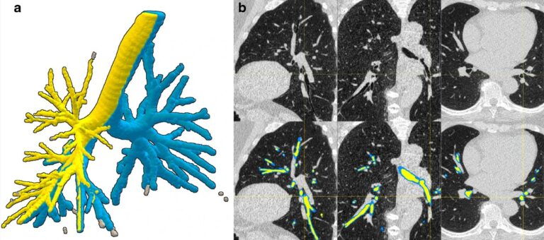
Reproducibility of a combined AI and optimal-surface graph-cut method to automate bronchial parameter extraction

The authors of this study evaluated the reproducibility of a deep learning and optimal-surface graph-cut method to automatically segment the airway lumen and wall, and calculate bronchial parameters. A deep-learning model was trained on 24 low-dose chest CT scans. The study demonstrated a comprehensive and fully automatic pipeline for bronchial parameter measurement on low-dose CT using open-source tools. Key points

Read More →

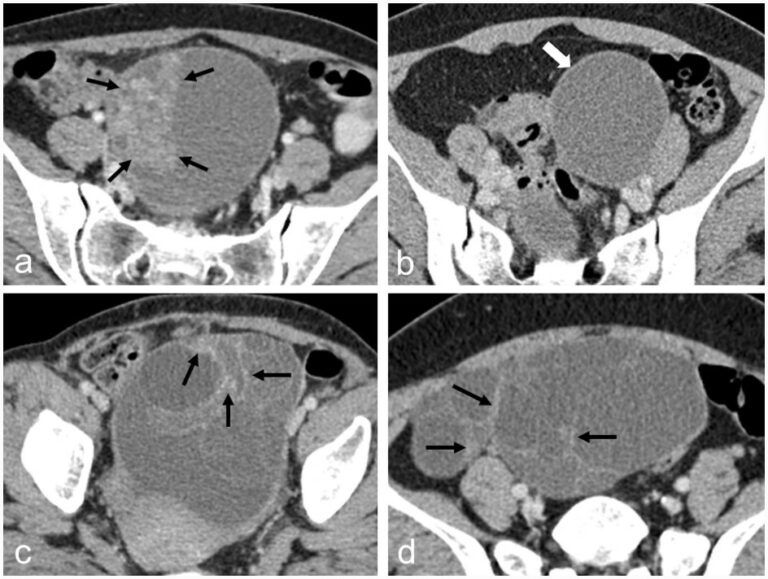
A novel AI model to distinguish benign from malignant ovarian tumors

The authors of this study developed a CT-based artificial intelligence model with the ability to differentiate between benign and malignant ovarian tumors, showing high accuracy and specificity. In coordination with less-experienced radiologists, the model helped in the performance of ovarian tumor assessment, with applications to provide better therapeutic strategies for patients with ovarian tumors. Key points CT-based radiomics and deep

Read More →

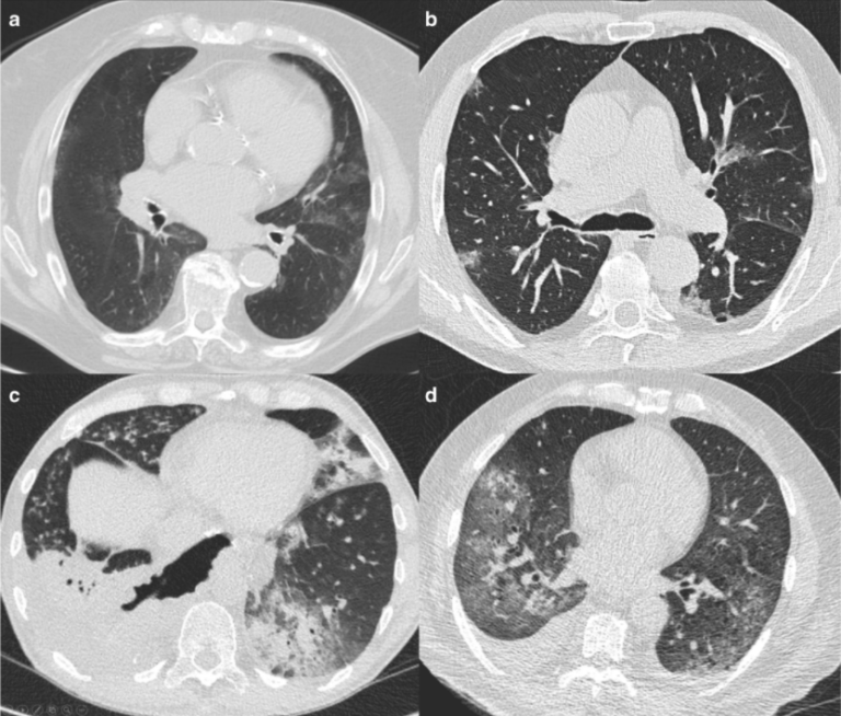
Commercial AI solutions in detecting COVID‐19 pneumonia in chest CT: not yet ready for clinical implementation?

Thinking back on the last two years, what were the dominant topics of discussion in radiology? Certainly, artificial intelligence (AI) in radiology has sparked a lot of interest and enthusiasm in radiology, and COVID-19, which was a topic nobody could avoid. So, it comes as no surprise that the combination of both topics – i.e. using AI to detect COVID

Read More →

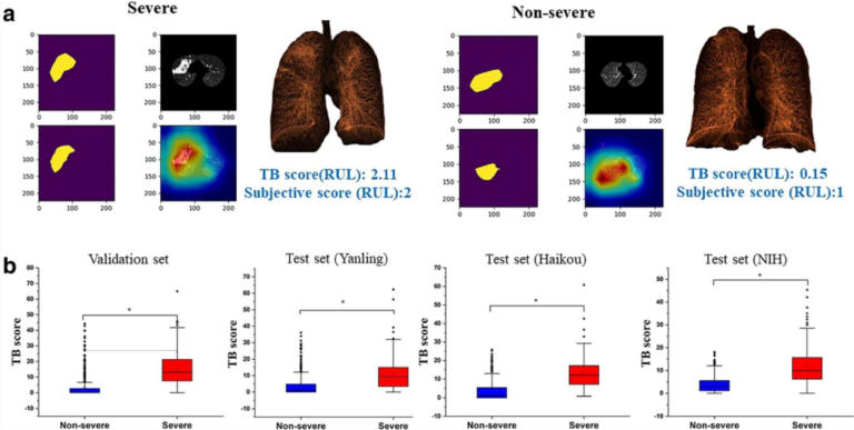
A fully automatic artificial intelligence–based CT image analysis system for accurate detection, diagnosis, and quantitative severity evaluation of pulmonary tuberculosis

The authors of this study aimed to develop an artificial intelligence (AI)-based fully automated CT image analysis system in order to detect and diagnose pulmonary tuberculosis (TB). This was achieved through the retrospective use of 892 chest CT scans from pathogen-confirmed TB patients. It was found that the end-to-end AI system based on chest CT is able to achieve human-level

Read More →

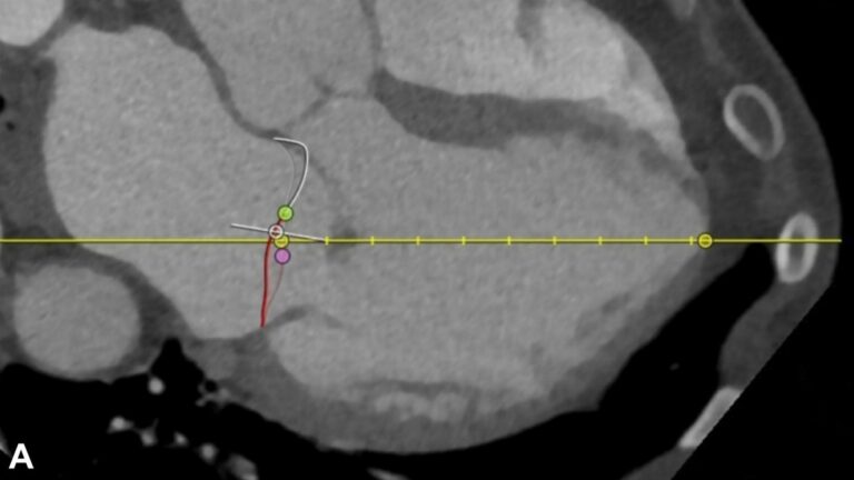
A fully automated software platform for structural mitral valve analysis

There has been great interest in the development and application of artificial intelligence (AI) in the field of medical imaging. AI has the potential to be useful in multiple arenas including the improvement of diagnostic accuracy as well as improving workflow and helping increase human efficiency in study interpretation. Cardiac imaging has been one focus of AI and has yielded

Read More →

Deep learning for automatic bowel-obstruction identification on abdominal CT

In our recent study, we developed an automated system for evaluating abdominal computed tomography (CT) scans to assist radiologists in managing their substantial workloads, thus improving patient outcomes. Our machine-learning model focuses on reliably identifying suspected bowel obstruction (BO) on abdominal CT scans. We used an internal dataset of 1,345 annotated CT scans, of which only 670 were re-annotated by

Read More →

Reproducibility of a combined AI and optimal-surface graph-cut method to automate bronchial parameter extraction

The authors of this study evaluated the reproducibility of a deep learning and optimal-surface graph-cut method to automatically segment the airway lumen and wall, and calculate bronchial parameters. A deep-learning model was trained on 24 low-dose chest CT scans. The study demonstrated a comprehensive and fully automatic pipeline for bronchial parameter measurement on low-dose CT using open-source tools. Key points

Read More →

A novel AI model to distinguish benign from malignant ovarian tumors

The authors of this study developed a CT-based artificial intelligence model with the ability to differentiate between benign and malignant ovarian tumors, showing high accuracy and specificity. In coordination with less-experienced radiologists, the model helped in the performance of ovarian tumor assessment, with applications to provide better therapeutic strategies for patients with ovarian tumors. Key points CT-based radiomics and deep

Read More →

Commercial AI solutions in detecting COVID‐19 pneumonia in chest CT: not yet ready for clinical implementation?

Thinking back on the last two years, what were the dominant topics of discussion in radiology? Certainly, artificial intelligence (AI) in radiology has sparked a lot of interest and enthusiasm in radiology, and COVID-19, which was a topic nobody could avoid. So, it comes as no surprise that the combination of both topics – i.e. using AI to detect COVID

Read More →

A fully automatic artificial intelligence–based CT image analysis system for accurate detection, diagnosis, and quantitative severity evaluation of pulmonary tuberculosis

The authors of this study aimed to develop an artificial intelligence (AI)-based fully automated CT image analysis system in order to detect and diagnose pulmonary tuberculosis (TB). This was achieved through the retrospective use of 892 chest CT scans from pathogen-confirmed TB patients. It was found that the end-to-end AI system based on chest CT is able to achieve human-level

Read More →

A fully automated software platform for structural mitral valve analysis

There has been great interest in the development and application of artificial intelligence (AI) in the field of medical imaging. AI has the potential to be useful in multiple arenas including the improvement of diagnostic accuracy as well as improving workflow and helping increase human efficiency in study interpretation. Cardiac imaging has been one focus of AI and has yielded

Read More →

Become A Member Today!

You will have access to a wide range of benefits that can help you advance your career and stay up-to-date with the latest developments in the field of radiology. These benefits include access to educational resources, networking opportunities with other professionals in the field, opportunities to participate in research projects and clinical trials, and access to the latest technologies and techniques. 

Check out our different membership options.

If you don’t find a fitting membership send us an email here.

Membership

for radiologists, radiology residents, professionals of allied sciences (including radiographers/radiological technologists, nuclear medicine physicians, medical physicists, and data scientists) & professionals of allied sciences in training residing within the boundaries of Europe

  • Reduced registration fees for ECR 1
  • Reduced fees for the European School of Radiology (ESOR) 2
  • Option to participate in the European Diploma. 3
  • Free electronic access to the journal European Radiology 
  • Content e-mails for all ESR journals4
  • Updates on offers & events through our newsletters
  • Exclusive access to the ESR feed in Juisci

€ 11 /year

Yes! That is less than €1 per month.

Free membership

for radiologists, radiology residents or professionals of allied sciences engaged in practice, teaching or research residing outside Europe as well as individual qualified professionals with an interest in radiology and medical imaging who do not fulfil individual or all requirements for any other ESR membership category & former full members who have retired from all clinical practice
  • Reduced registration fees for ECR 1
  • Option to participate in the European Diploma. 3
  • Free electronic access to the journal European Radiology
  • Content e-mails for all ESR journals 4
  • Updates on offers & events through our newsletters
  • Exclusive access to the ESR feed in Juisci

€ 0

The best things in life are free.

ESR Friends

For students, company representatives or hospital managers etc.

  • Content e-mails for all 3 ESR journals 4
  • Updates on offers & events through our newsletters

€ 0

Friendship doesn’t cost a thing.

The membership type best fitting for you will be selected automatically during the application process.

Footnotes:

01

Reduced registration fees for ECR 2025:
Provided that ESR 2024 membership is activated and approved by August 31, 2024.

Reduced registration fees for ECR 2026:
Provided that ESR 2025 membership is activated and approved by August 31, 2025.

02
Not all activities included
03
Examination based on the ESR European Training Curriculum (radiologists or radiology residents).
04
European Radiology, Insights into Imaging, European Radiology Experimental.