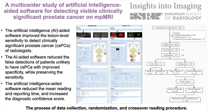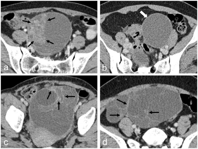
A novel AI model to distinguish benign from malignant ovarian tumors
The authors of this study developed a CT-based artificial intelligence model with the ability to differentiate between benign and malignant ovarian tumors, showing high accuracy and specificity. In coordination with less-experienced radiologists, the model helped in the performance of ovarian tumor assessment, with applications to provide better therapeutic strategies for patients with ovarian tumors. Key points CT-based radiomics and deep