Fully automated pelvic bone segmentation in multiparametric MRI using a 3D convolutional neural network
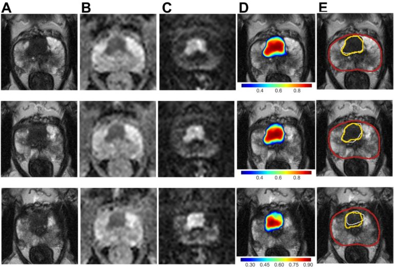
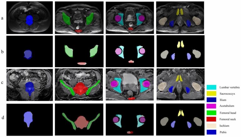
The accurate skeleton segmentation with their semantic labels represents the initial step to achieve accurate prostate cancer bone metastases detection on DWI and ADC images. Several studies have reported convolutional neural networks (CNNs) for the segmentation of normal bone structures on CT images and bone scans; however, only a few studies on automatic segmentation of normal bone structures on MR