Improved long-term prognostic value of coronary CT angiography-derived plaque measures and clinical parameters on adverse cardiac outcome using machine learning
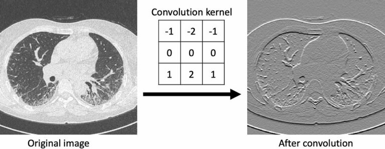
In recent years, artificial intelligence (AI) and, in particular, the application of machine learning (ML) algorithms, have become a new cornerstone in cardiovascular imaging with improved decision pathways, risk stratification, and outcome prediction in a more objective, reproducible, and rational manner. The integration of ML in daily routine clinical practice may hold potential to improve imaging workflow and to promote