prostatic neoplasms

Welcome to the blog on Artificial Intelligence of
the European Society of Radiology

This blog aims at bringing educational and critical perspectives on AI to readers. It should help imaging professionals to learn and keep up to date with the technologies being developed in this rapidly evolving field.

Most used hashtags:

Latest posts

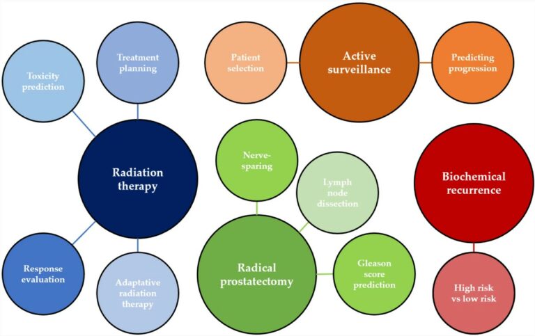
Beyond diagnosis: is there a role for radiomics in prostate cancer management?

At present, therapeutic and prognostic recommendations for prostate cancer (PCa) predominantly hinge on risk-stratification tools that are built upon clinical parameters. Recent evidence indicates that incorporating imaging can enhance the precision of prognostic models based on clinical factors. However, challenges like subjective interpretation, variability in image analysis, and the absence of reliable quantitative measures need to be overcome to fully

Read More →

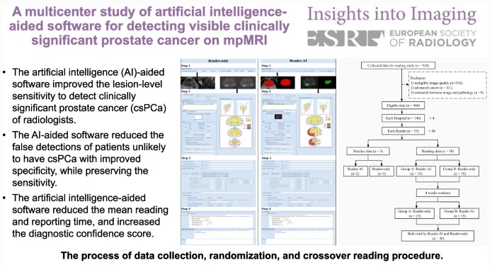
AI-aided software for detecting visible clinically significant prostate cancer on mpMRI

This study seeks to determine if artificial intelligence (AI)-based software can improve radiologists’ performance when detecting clinically significant prostate cancer. Sixteen radiologists from four hospitals participated and were assigned 30 cases, half without AI and half with AI. The authors determined that the AI software improves the performance of radiologists by reducing false positive detection of prostate cancer patients while

Read More →

AI for prostate MRI: open datasets, available applications, and grand challenges

This narrative review provides an overview of the current state-of-the-art artificial intelligence (AI) applications for prostate MRI by focusing on open datasets, commercially and publically available AI systems, and challenges. The authors state that large amounts of research are still required in order to successfully utilize AI in the whole prostate pathway. Due to the rapidly growing field, continuous up-to-date

Read More →

Tasks for AI in prostate MRI

The authors of this narrative review aimed to introduce quality metrics for emerging artificial intelligence (AI) papers, such as the Checklist for Artificial Intelligence in Medical Imaging (CLAIM) and Field-Weighted Citation Impact (FWCI). Furthermore, the study dives into some of the top AI models for segmentation, detection, and classification, while concluding that prospective studies with multi-center design will need to

Read More →

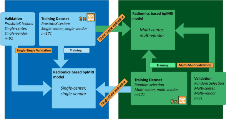
Single-center versus multi-center biparametric MRI radiomics approach

In this study, the authors’ aim was to investigate a previously developed radiomics-based biparametric magnetic resonance imaging (bpMRI) approach used for the discrimination of clinically significant peripheral zone prostate cancer (PZ csPCa) through the use of multi-center, multi-vendor (McMv) and single-center, single-vendor (ScSv) datasets. Using these datasets, the authors were able to determine that a single-center trained radiomics-based bpMRI model

Read More →

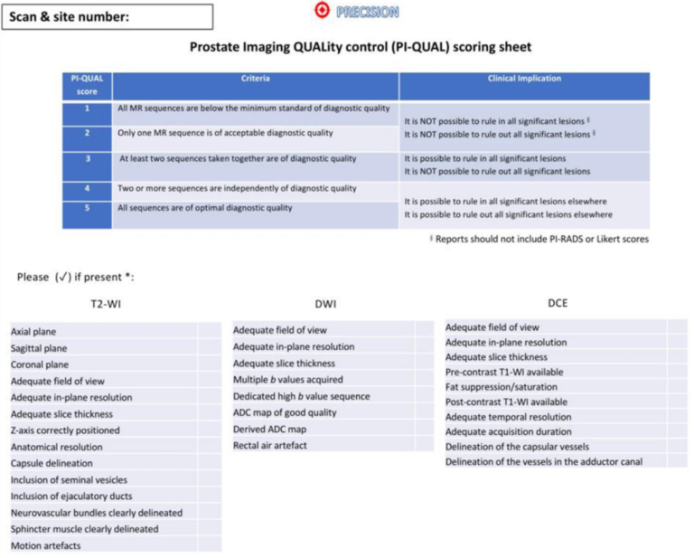
Multiparametric prostate MRI quality assessment using a PI-QUAL software program

We know that the technical requirements for the acquisition of multiparametric MRI of the prostate have been clearly outlined in the PI-RADS guidelines, but there is still huge variability in image quality among centres across the world. The Prostate Imaging Quality (PI-QUAL) score and its dedicated scoring sheet represent the first attempt to standardise image quality, as they take into

Read More →

Multiparametric MRI and auto-fixed volume of interest-based radiomics signature for clinically significant peripheral zone prostate cancer

The purpose of this study was to create a radiomics approach based on multiparametric MRI (mpMRI) features that were extracted from an auto-fixed volume of interest (VOI) that quantifies the phenotype of clinically significant peripheral zone prostate cancer (pCA). The study included 206 patients and the authors concluded that the developed radiomics model that extracts mpMRI features with an auto-fixed

Read More →

Beyond diagnosis: is there a role for radiomics in prostate cancer management?

At present, therapeutic and prognostic recommendations for prostate cancer (PCa) predominantly hinge on risk-stratification tools that are built upon clinical parameters. Recent evidence indicates that incorporating imaging can enhance the precision of prognostic models based on clinical factors. However, challenges like subjective interpretation, variability in image analysis, and the absence of reliable quantitative measures need to be overcome to fully

Read More →

AI-aided software for detecting visible clinically significant prostate cancer on mpMRI

This study seeks to determine if artificial intelligence (AI)-based software can improve radiologists’ performance when detecting clinically significant prostate cancer. Sixteen radiologists from four hospitals participated and were assigned 30 cases, half without AI and half with AI. The authors determined that the AI software improves the performance of radiologists by reducing false positive detection of prostate cancer patients while

Read More →

AI for prostate MRI: open datasets, available applications, and grand challenges

This narrative review provides an overview of the current state-of-the-art artificial intelligence (AI) applications for prostate MRI by focusing on open datasets, commercially and publically available AI systems, and challenges. The authors state that large amounts of research are still required in order to successfully utilize AI in the whole prostate pathway. Due to the rapidly growing field, continuous up-to-date

Read More →

Tasks for AI in prostate MRI

The authors of this narrative review aimed to introduce quality metrics for emerging artificial intelligence (AI) papers, such as the Checklist for Artificial Intelligence in Medical Imaging (CLAIM) and Field-Weighted Citation Impact (FWCI). Furthermore, the study dives into some of the top AI models for segmentation, detection, and classification, while concluding that prospective studies with multi-center design will need to

Read More →

Single-center versus multi-center biparametric MRI radiomics approach

In this study, the authors’ aim was to investigate a previously developed radiomics-based biparametric magnetic resonance imaging (bpMRI) approach used for the discrimination of clinically significant peripheral zone prostate cancer (PZ csPCa) through the use of multi-center, multi-vendor (McMv) and single-center, single-vendor (ScSv) datasets. Using these datasets, the authors were able to determine that a single-center trained radiomics-based bpMRI model

Read More →

Multiparametric prostate MRI quality assessment using a PI-QUAL software program

We know that the technical requirements for the acquisition of multiparametric MRI of the prostate have been clearly outlined in the PI-RADS guidelines, but there is still huge variability in image quality among centres across the world. The Prostate Imaging Quality (PI-QUAL) score and its dedicated scoring sheet represent the first attempt to standardise image quality, as they take into

Read More →

Multiparametric MRI and auto-fixed volume of interest-based radiomics signature for clinically significant peripheral zone prostate cancer

The purpose of this study was to create a radiomics approach based on multiparametric MRI (mpMRI) features that were extracted from an auto-fixed volume of interest (VOI) that quantifies the phenotype of clinically significant peripheral zone prostate cancer (pCA). The study included 206 patients and the authors concluded that the developed radiomics model that extracts mpMRI features with an auto-fixed

Read More →

Become A Member Today!

You will have access to a wide range of benefits that can help you advance your career and stay up-to-date with the latest developments in the field of radiology. These benefits include access to educational resources, networking opportunities with other professionals in the field, opportunities to participate in research projects and clinical trials, and access to the latest technologies and techniques. 

Check out our different membership options.

If you don’t find a fitting membership send us an email here.

Membership

for radiologists, radiology residents, professionals of allied sciences (including radiographers/radiological technologists, nuclear medicine physicians, medical physicists, and data scientists) & professionals of allied sciences in training residing within the boundaries of Europe

  • Reduced registration fees for ECR 1
  • Reduced fees for the European School of Radiology (ESOR) 2
  • Option to participate in the European Diploma. 3
  • Free electronic access to the journal European Radiology 
  • Content e-mails for all ESR journals4
  • Updates on offers & events through our newsletters
  • Exclusive access to the ESR feed in Juisci

€ 11 /year

Yes! That is less than €1 per month.

Free membership

for radiologists, radiology residents or professionals of allied sciences engaged in practice, teaching or research residing outside Europe as well as individual qualified professionals with an interest in radiology and medical imaging who do not fulfil individual or all requirements for any other ESR membership category & former full members who have retired from all clinical practice
  • Reduced registration fees for ECR 1
  • Option to participate in the European Diploma. 3
  • Free electronic access to the journal European Radiology
  • Content e-mails for all ESR journals 4
  • Updates on offers & events through our newsletters
  • Exclusive access to the ESR feed in Juisci

€ 0

The best things in life are free.

ESR Friends

For students, company representatives or hospital managers etc.

  • Content e-mails for all 3 ESR journals 4
  • Updates on offers & events through our newsletters

€ 0

Friendship doesn’t cost a thing.

The membership type best fitting for you will be selected automatically during the application process.

Footnotes:

01

Reduced registration fees for ECR 2025:
Provided that ESR 2024 membership is activated and approved by August 31, 2024.

Reduced registration fees for ECR 2026:
Provided that ESR 2025 membership is activated and approved by August 31, 2025.

02
Not all activities included
03
Examination based on the ESR European Training Curriculum (radiologists or radiology residents).
04
European Radiology, Insights into Imaging, European Radiology Experimental.