radiomics

Welcome to the blog on Artificial Intelligence of
the European Society of Radiology

This blog aims at bringing educational and critical perspectives on AI to readers. It should help imaging professionals to learn and keep up to date with the technologies being developed in this rapidly evolving field.

Most used hashtags:

Latest posts

Radiomic feature reproducibility in contrast-enhanced CT of the pancreas is affected by variabilities in scan parameters and manual segmentation

The goal of this study was to measure the reproducibility of radiomic features in pancreatic parenchyma and ductal adenocarcinomas in patients who underwent consecutive contrast-enhanced computed tomography (CECT) scans. A majority of features were not reproducible (as defined by a concordance correlation coefficient of greater than 0.9) when comparing their values across consecutive CECTs obtained within a two week period.

Read More →

Radiomics machine-learning signature for diagnosis of hepatocellular carcinoma in cirrhotic patients with indeterminate liver nodules

This article sets out to determine whether machine learning can be used to train and calibrate the signature for diagnosing hepatocellular carcinoma in cirrhotic patients with indeterminate liver nodules. The authors proved that artificial intelligence could enhance clinicians’ decision and reduce the rate of cirrhotic patients requiring liver biopsy. Key points In cirrhotic patients with visually indeterminate liver nodules, expert

Read More →

Breast cancer Ki-67 expression prediction by digital breast tomosynthesis radiomics features

Despite the encouraging results, more studies are needed in order to further evaluate these preliminary findings and to find to what extent radiomics and AI approaches can be integrated in clinical practice in a useful and reliable strategy [1]. I think that several issues reduce the application of radiomics approaches in clinical practice: the lack of knowledge of its basic

Read More →

Why radiomics research does not translate to clinical practice: evaluation of literature using RQS and TRIPOD

Over the last few years, the number of studies published using quantitative imaging biomarkers to classify or predict pathologies has steadily increased. As of today, a quick PubMed search for radiomics, imaging biomarkers or radiogenomics reveals well over 4,000 articles. However, somewhat surprisingly, given this amount of published research, outside of academic literature there is no widespread clinical application of

Read More →

MRI radiomics in categorizing ovarian masses and predicting clinical outcome: a preliminary study

This study aimed to assess whether MRI radiomics can categorize ovarian masses and to determine the association between MRI radiomics and survival among ovarian epithelial cancer patients. The authors evaluated the diagnostic performance of the signatures derived from MRI radiomics in 286 patients with proven adnexal tumor. The study results suggest a correlation between radiomics features extracted from MRI and

Read More →

What the radiologist should know about artificial intelligence – an ESR white paper

Did you ever get lost between all the buzz words floating around like artificial intelligence, imaging informatics, radiomics, imaging biobanks, neural networks, clinical decision support, etc.? And what are the ethical and medico-legal implications of all these new developments? Then you may want to read the ESR white paper that was recently published by the ESR’s eHealth and Informatics subcommittee

Read More →

How machine learning-based high-dimensional CT texture analysis is influenced by segmentation margin

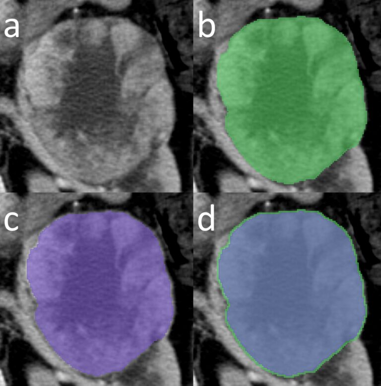
Radiomic workflows include various challenging steps. One of the most demanding steps in radiomics is the segmentation process. Particularly for the renal cell carcinomas, most of the studies used manual tumour contour delineation. In this work, our group wanted to perform an experiment by changing the segmentation margin a little bit, that is, just 2 mm, to see what happens

Read More →

Radiomics: a critical step towards integrated healthcare

This article aims to bring together the various technological developments that have taken place in medical imaging analysis and highlight a potential path for the future. While the term “medical image analysis” has classically referred to radiological images (CT, MRI, PET, etc.), we must also remember that digitalization occurred much earlier in other diagnostic disciplines like pathology (with pathomics) and

Read More →

Radiomics: the facts and the challenges of image analysis

Radiomics is a complex multi-step process that can be considered as part of the more complex world of Artifical Intelligence (AI). The aim of radiomics is aiding clinical decision-making and outcome prediction for more personalized medicine. Each step of the radiomics process brings challenges that have to be considered; for example, segmentation is challenging because of reproducibility issues. Indeed, there

Read More →

Comparing computer- and human-extracted imaging phenotypes

This retrospective study sought to investigate if computer-extracted magnetic resonance imaging (MRI) phenotypes of breast cancer could replicate human-extracted size and Breast Imaging-Reporting and Data System (BI-RADS) imaging phenotypes using MRI data from The Cancer Genome Atlas (TCGA) project of the National Cancer Institute. Upon obtaining the results, it was possible to conclude that quantitative radiomics of breast cancer may replicate human-extracted tumour size

Read More →

Radiomic feature reproducibility in contrast-enhanced CT of the pancreas is affected by variabilities in scan parameters and manual segmentation

The goal of this study was to measure the reproducibility of radiomic features in pancreatic parenchyma and ductal adenocarcinomas in patients who underwent consecutive contrast-enhanced computed tomography (CECT) scans. A majority of features were not reproducible (as defined by a concordance correlation coefficient of greater than 0.9) when comparing their values across consecutive CECTs obtained within a two week period.

Read More →

Radiomics machine-learning signature for diagnosis of hepatocellular carcinoma in cirrhotic patients with indeterminate liver nodules

This article sets out to determine whether machine learning can be used to train and calibrate the signature for diagnosing hepatocellular carcinoma in cirrhotic patients with indeterminate liver nodules. The authors proved that artificial intelligence could enhance clinicians’ decision and reduce the rate of cirrhotic patients requiring liver biopsy. Key points In cirrhotic patients with visually indeterminate liver nodules, expert

Read More →

Breast cancer Ki-67 expression prediction by digital breast tomosynthesis radiomics features

Despite the encouraging results, more studies are needed in order to further evaluate these preliminary findings and to find to what extent radiomics and AI approaches can be integrated in clinical practice in a useful and reliable strategy [1]. I think that several issues reduce the application of radiomics approaches in clinical practice: the lack of knowledge of its basic

Read More →

Why radiomics research does not translate to clinical practice: evaluation of literature using RQS and TRIPOD

Over the last few years, the number of studies published using quantitative imaging biomarkers to classify or predict pathologies has steadily increased. As of today, a quick PubMed search for radiomics, imaging biomarkers or radiogenomics reveals well over 4,000 articles. However, somewhat surprisingly, given this amount of published research, outside of academic literature there is no widespread clinical application of

Read More →

MRI radiomics in categorizing ovarian masses and predicting clinical outcome: a preliminary study

This study aimed to assess whether MRI radiomics can categorize ovarian masses and to determine the association between MRI radiomics and survival among ovarian epithelial cancer patients. The authors evaluated the diagnostic performance of the signatures derived from MRI radiomics in 286 patients with proven adnexal tumor. The study results suggest a correlation between radiomics features extracted from MRI and

Read More →

What the radiologist should know about artificial intelligence – an ESR white paper

Did you ever get lost between all the buzz words floating around like artificial intelligence, imaging informatics, radiomics, imaging biobanks, neural networks, clinical decision support, etc.? And what are the ethical and medico-legal implications of all these new developments? Then you may want to read the ESR white paper that was recently published by the ESR’s eHealth and Informatics subcommittee

Read More →

How machine learning-based high-dimensional CT texture analysis is influenced by segmentation margin

Radiomic workflows include various challenging steps. One of the most demanding steps in radiomics is the segmentation process. Particularly for the renal cell carcinomas, most of the studies used manual tumour contour delineation. In this work, our group wanted to perform an experiment by changing the segmentation margin a little bit, that is, just 2 mm, to see what happens

Read More →

Radiomics: a critical step towards integrated healthcare

This article aims to bring together the various technological developments that have taken place in medical imaging analysis and highlight a potential path for the future. While the term “medical image analysis” has classically referred to radiological images (CT, MRI, PET, etc.), we must also remember that digitalization occurred much earlier in other diagnostic disciplines like pathology (with pathomics) and

Read More →

Radiomics: the facts and the challenges of image analysis

Radiomics is a complex multi-step process that can be considered as part of the more complex world of Artifical Intelligence (AI). The aim of radiomics is aiding clinical decision-making and outcome prediction for more personalized medicine. Each step of the radiomics process brings challenges that have to be considered; for example, segmentation is challenging because of reproducibility issues. Indeed, there

Read More →

Comparing computer- and human-extracted imaging phenotypes

This retrospective study sought to investigate if computer-extracted magnetic resonance imaging (MRI) phenotypes of breast cancer could replicate human-extracted size and Breast Imaging-Reporting and Data System (BI-RADS) imaging phenotypes using MRI data from The Cancer Genome Atlas (TCGA) project of the National Cancer Institute. Upon obtaining the results, it was possible to conclude that quantitative radiomics of breast cancer may replicate human-extracted tumour size

Read More →

Become A Member Today!

You will have access to a wide range of benefits that can help you advance your career and stay up-to-date with the latest developments in the field of radiology. These benefits include access to educational resources, networking opportunities with other professionals in the field, opportunities to participate in research projects and clinical trials, and access to the latest technologies and techniques. 

Check out our different membership options.

If you don’t find a fitting membership send us an email here.

Membership

for radiologists, radiology residents, professionals of allied sciences (including radiographers/radiological technologists, nuclear medicine physicians, medical physicists, and data scientists) & professionals of allied sciences in training residing within the boundaries of Europe

  • Reduced registration fees for ECR 1
  • Reduced fees for the European School of Radiology (ESOR) 2
  • Option to participate in the European Diploma. 3
  • Free electronic access to the journal European Radiology 
  • Content e-mails for all ESR journals4
  • Updates on offers & events through our newsletters
  • Exclusive access to the ESR feed in Juisci

€ 11 /year

Yes! That is less than €1 per month.

Free membership

for radiologists, radiology residents or professionals of allied sciences engaged in practice, teaching or research residing outside Europe as well as individual qualified professionals with an interest in radiology and medical imaging who do not fulfil individual or all requirements for any other ESR membership category & former full members who have retired from all clinical practice
  • Reduced registration fees for ECR 1
  • Option to participate in the European Diploma. 3
  • Free electronic access to the journal European Radiology
  • Content e-mails for all ESR journals 4
  • Updates on offers & events through our newsletters
  • Exclusive access to the ESR feed in Juisci

€ 0

The best things in life are free.

ESR Friends

For students, company representatives or hospital managers etc.

  • Content e-mails for all 3 ESR journals 4
  • Updates on offers & events through our newsletters

€ 0

Friendship doesn’t cost a thing.

The membership type best fitting for you will be selected automatically during the application process.

Footnotes:

01

Reduced registration fees for ECR 2025:
Provided that ESR 2024 membership is activated and approved by August 31, 2024.

02
Not all activities included
03
Examination based on the ESR European Training Curriculum (radiologists or radiology residents).
04
European Radiology, Insights into Imaging, European Radiology Experimental.