AI Blog

Welcome to the blog on Artificial Intelligence of
the European Society of Radiology

This blog aims at bringing educational and critical perspectives on AI to readers. It should help imaging professionals to learn and keep up to date with the technologies being developed in this rapidly evolving field.

Most used hashtags:

Latest posts

Evaluation of a CTA-based convolutional neural network for infarct volume prediction in anterior cerebral circulation ischaemic stroke

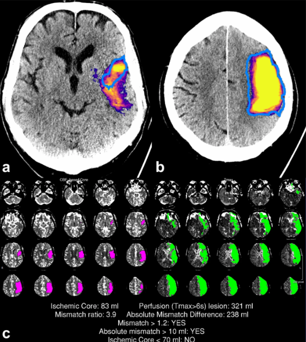
The authors of this study aimed to determine the efficacy of a convolutional neural network (CNN) in final infarct volume prediction from computed tomography angiography (CTA), subsequently comparing the results to a CT perfusion (CTP)-based commercially available software. The stroke cases treated with thrombolytic therapy or receiving supportive care were retrospectively selected by the authors. The study found that a

Read More →

CT and MRI radiomics of bone and soft-tissue sarcomas: a systematic review of reproducibility and validation strategies

The authors of this study aimed to systematically review radiomic feature reproducibility and predictive model validation strategies in studies that deal with CT and MRI radiomics of bone and soft-tissue sarcomas. The review consisted of 278 papers, forty-nine of which were published between 2008 and 2020. The authors found that the issues of radiomic feature reproducibility and model validation varied

Read More →

Machine learning automatically detects COVID-19 using chest CTs in a large multicenter cohort

This retrospective, multi-institutional study investigated machine learning classifiers and interpretable models using chest CT for the detection of COVID-19 and to differentiate this from types of pneumonia, interstitial lung disease (ILD), and normal CTs. The study included 2,446 chest CTs from across 16 different institutions and the authors’ method was found to accurately differentiate COVID-19 from other types of pneumonia,

Read More →

Automatic prediction of left cardiac chamber enlargement from chest radiographs using convolutional neural network

The aim of this study was to develop deep learning-based cardiac chamber enlargement-detection algorithms for left atrial (DLCE-LAE) and ventricular enlargement (DLCE-LVE) on chest radiographs. The authors determined that the DLCE-LAE was able to outperform and improve the performance of cardiothoracic radiologists in the detection of LAE, while also showing promise in screening individuals with moderate-to-severe LAE in a healthcare

Read More →

Artificial intelligence in medical imaging practice in Africa: a qualitative content analysis study of radiographers’ perspectives

The aim of this study was to qualitatively explore the perception of radiographers in relation to the integration and acceptance of artificial intelligence (AI) in medical imaging practice on the continent of Africa. Participants of this study consisted solely of radiographers working in Africa between March and August 2020. The study demonstrated a positive outlook regarding AI in relation to

Read More →

Impact of artificial intelligence support on accuracy and reading time in breast tomosynthesis image interpretation: a multi-reader multi-case study

In this multi-reader, multi-case study, the authors aimed to investigate whether an artificial intelligence (AI) support system could help to increase the accuracy of breast radiologists reading wide-angle digital breast tomosynthesis (DBT). The study was performed using 240 bilateral DBT exams, with the exams interpreted by 18 radiologists with and without AI support. The authors found that the radiologists improved

Read More →

Proposing a deep learning-based method for improving the diagnostic certainty of pulmonary nodules in CT scan of chest

The aim of this study was to compare the performance of a deep learning (DL)-based method used for diagnosing pulmonary nodules compared with the diagnostic approach of the radiologist in computed tomography (CT) of the chest. The authors included a total of 150 pathologically confirmed pulmonary nodules that were assessed and reported by radiologists. The study found that the DL-based

Read More →

Radiomics analysis of contrast-enhanced CT for classification of hepatic focal lesions in colorectal cancer patients: its limitations compared to radiologists

The authors of this retrospective study aimed to evaluate the diagnostic performance of a radiomics model in order to classify hepatic cyst, hemangioma, and metastasis in patients who have been diagnosed with colorectal cancer (CRC) from portal-phase abdominopelvic CT images. The study found that, although inferior to radiologists, the radiomics model was able to achieve substantial diagnostic performance when differentiating

Read More →

ESUR/ESUI position paper: developing artificial intelligence for precision diagnosis of prostate cancer using magnetic resonance imaging

The clinical promise of artificial intelligence (AI) in prostate cancer diagnosis has yet to materialize. Any AI application must reach an appropriate level of maturity and robustness for such developments to be accepted by its intended users. Our position paper, “Development of Artificial Intelligence for Precision Diagnosis of Prostate Cancer Using MRI”, co-authored by experts from ESUR and ESUI, elaborated

Read More →

Differential diagnosis of benign and malignant vertebral fracture on CT using deep learning

The purpose of this study was to evaluate the performance of deep learning using ResNet50 in the differentiation of benign and malignant vertebral fracture on computed tomography (CT). The study used a dataset of 433 patients, which was retrospectively selected from the authors’ spinal CT image database. The authors concluded that ResNet50 achieved good accuracy, which can be further improved

Read More →

Become A Member Today!

You will have access to a wide range of benefits that can help you advance your career and stay up-to-date with the latest developments in the field of radiology. These benefits include access to educational resources, networking opportunities with other professionals in the field, opportunities to participate in research projects and clinical trials, and access to the latest technologies and techniques. 

Check out our different membership options.

If you don’t find a fitting membership send us an email here.

Membership

for radiologists, radiology residents, professionals of allied sciences (including radiographers/radiological technologists, nuclear medicine physicians, medical physicists, and data scientists) & professionals of allied sciences in training residing within the boundaries of Europe

  • Reduced registration fees for ECR 1
  • Reduced fees for the European School of Radiology (ESOR) 2
  • Option to participate in the European Diploma. 3
  • Free electronic access to the journal European Radiology 4
  • Content e-mails for all ESR journals
  • Updates on offers & events through our newsletters
  • Exclusive access to the ESR feed in Juisci

€ 11 /year

Yes! That is less than €1 per month.

Free membership

for radiologists, radiology residents or professionals of allied sciences engaged in practice, teaching or research residing outside Europe as well as individual qualified professionals with an interest in radiology and medical imaging who do not fulfil individual or all requirements for any other ESR membership category & former full members who have retired from all clinical practice
  • Reduced registration fees for ECR 1
  • Option to participate in the European Diploma. 3
  • Free electronic access to the journal European Radiology
  • Content e-mails for all 3 ESR journals 4
  • Updates on offers & events through our newsletters
  • Exclusive access to the ESR feed in Juisci

€ 0

The best things in life are free.

ESR Friends

For students, company representatives or hospital managers etc.

  • Content e-mails for all 3 ESR journals 4
  • Updates on offers & events through our newsletters

€ 0

Friendship doesn’t cost a thing.

The membership type best fitting for you will be selected automatically during the application process.

Footnotes:

01

Reduced registration fees for ECR 2025:
Provided that ESR 2024 membership is activated and approved by August 31, 2024.

02
Not all activities included
03
Examination based on the ESR European Training Curriculum (radiologists or radiology residents).
04
European Radiology, Insights into Imaging, European Radiology Experimental.