AI Blog

Welcome to the blog on Artificial Intelligence of
the European Society of Radiology

This blog aims at bringing educational and critical perspectives on AI to readers. It should help imaging professionals to learn and keep up to date with the technologies being developed in this rapidly evolving field.

Most used hashtags:

Latest posts

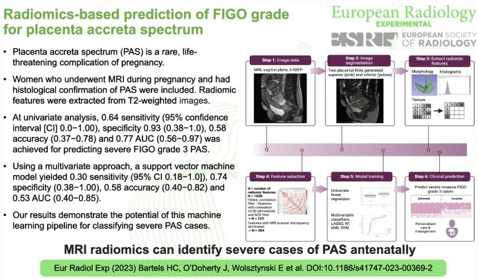
Radiomics-based prediction of FIGO grade for placenta accreta spectrum

Placenta Accreta Spectrum (PAS) is a serious, life-threatening pregnancy complication. Although rare, as more women are giving birth by Caesarean section, PAS is becoming more common. The most important factor in improving outcomes for mothers and babies is the detection of PAS during pregnancy to ensure the appropriate multi-disciplinary team care is implemented for the pregnancy and birth. However, up

Read More →

Can radiomics push the limits of current IPMN malignancy assessment and help avoid unnecessary resection?

This commentary dives into an article published in August 2023 in European Radiology entitled “Radiomics model versus 2017 revised international consensus guidelines for predicting malignant intraductal papillary mucinous neoplasms” (Doo Young Lee et al.), which compared the diagnostic performance of the CT radiomics model with the 2017 international consensus Fukuoka guideline for predicting malignant intraductal papillary mucinous neoplasms (IMPNs). The

Read More →

Self-reporting with checklists in AI research on medical imaging

This study aimed to evaluate the usage of the Checklist for Artificial Intelligence in Medical Imaging (CLAIM), a well-known and widely adopted checklist in the radiological community, for self-reporting through a systematic analysis of its citations. The authors used three databases (Google Scholar, Web of Science, and Scopus) and identified nearly 400 unique citations across 118 papers, of which only

Read More →

Ovarian cancer beyond imaging: integration of AI and multiomics biomarkers

Ovarian cancer, often referred to as the “silent killer”, tends to show inconspicuous symptoms in the early stages, making timely diagnosis difficult. Detecting ovarian cancer at an early stage significantly increases the chances of effective treatment and therefore the chances of survival. This review study has shown that AI-based tools that rely on the integration of multi-omics data perform better

Read More →

Label-set impact on deep learning-based prostate segmentation on MRI

This study delves into a less-explored territory in automatic prostate segmentation: label-set selection. Recognizing the emphasis on dataset selection in segmentation model training, we thought it crucial to investigate the impact of the labels, i.e. the manual segmentations, on model performance. Although label sets are often considered the gold standard, as they are provided by highly trained professionals, disparities emerge

Read More →

External validation, radiological evaluation, and development of DL lung segmentation in chest CT

The authors of this study developed a 3D nnU-Net-based model for automatic lung segmentation in computed tomography pulmonary angiography (CTPA) imaging that was found to be highly accurate, clinically evaluated, and externally tested in patient cohorts with a spread of lung disease. Key points Article: External validation, radiological evaluation, and development of deep learning automatic lung segmentation in contrast-enhanced chest CT

Read More →

ChatGPT makes medicine easy to swallow

Just over a year ago, the release of ChatGPT marked a turning point in AI and language processing, sparking widespread excitement for Large Language Models (LLMs) for diverse use cases across various domains. Back then, we noticed friends using ChatGPT for medical text simplification, yet, as medical laypersons, they couldn’t verify the accuracy of the simplified text. Anticipating its imminent adoption by patients,

Read More →

Core and penumbra estimation using deep learning-based AIF in association with clinical measures in computed tomography perfusion (CTP)

In this study, the utilization of a convolutional neural network (CNN)-based arterial input function (AIF) and whether it improves the volumetric estimation of core penumbra was investigated. The authors included 160 acute ischemic stroke patients for this study, who underwent CTP imaging, National Institutes of Health Stroke Scale (NIHSS), and Alberta Stroke Programme Early CT Score (APSECTS) grading. They were

Read More →

Revolutionising paediatric radiology: the future impact of AI

There remains a hesitancy regarding the adoption of artificial intelligence (AI) within the subspecialties of radiology and whether it has a fixed role in radiology’s future; this is especially true in paediatric radiology. Factors such as a lack of trust in AI applications and a lack of IT infrastructure in many hospitals, among other things, contribute largely to this hesitancy. This

Read More →

Evaluating AI for Clinical Decision-Making: Lessons Learned from a Study of ChatGPT’s Referral Reliability

In our recent study published in European Radiology, we evaluated the reliability of ChatGPT – an AI system developed by OpenAI – as a referral tool for imaging tests, compared to ESR iGuide, a clinical decision support system (CDSS) developed by the European Society of Radiology in cooperation with the American College of Radiology. Four experts served as our ground

Read More →

Become A Member Today!

You will have access to a wide range of benefits that can help you advance your career and stay up-to-date with the latest developments in the field of radiology. These benefits include access to educational resources, networking opportunities with other professionals in the field, opportunities to participate in research projects and clinical trials, and access to the latest technologies and techniques. 

Check out our different membership options.

If you don’t find a fitting membership send us an email here.

Membership

for radiologists, radiology residents, professionals of allied sciences (including radiographers/radiological technologists, nuclear medicine physicians, medical physicists, and data scientists) & professionals of allied sciences in training residing within the boundaries of Europe

  • Reduced registration fees for ECR 1
  • Reduced fees for the European School of Radiology (ESOR) 2
  • Exclusive option to participate in the European Diploma. 3
  • Free electronic access to the journal European Radiology 4
  • Content e-mails for all ESR journals
  • Updates on offers & events through our newsletters
  • Exclusive access to the ESR feed in Juisci

€ 11 /year

Yes! That is less than €1 per month.

Free membership

for radiologists, radiology residents or professionals of allied sciences engaged in practice, teaching or research residing outside Europe as well as individual qualified professionals with an interest in radiology and medical imaging who do not fulfil individual or all requirements for any other ESR membership category & former full members who have retired from all clinical practice
  • Reduced registration fees for ECR 1
  • Free electronic access to the journal European Radiology
  • Content e-mails for all 3 ESR journals 4
  • Updates on offers & events through our newsletters
  • Exclusive access to the ESR feed in Juisci

€ 0

The best things in life are free.

ESR Friends

For students, company representatives or hospital managers etc.

  • Content e-mails for all 3 ESR journals 4
  • Updates on offers & events through our newsletters

€ 0

Friendship doesn’t cost a thing.

The membership type best fitting for you will be selected automatically during the application process.

Footnotes:

01

Reduced registration fees for ECR 2024:
Provided that ESR 2023 membership is activated and approved by August 31, 2023.

Reduced registration fees for ECR 2025:
Provided that ESR 2024 membership is activated and approved by August 31, 2024.

02
Not all activities included
03
Examination based on the ESR European Training Curriculum (radiologists or radiology residents).
04
European Radiology, Insights into Imaging, European Radiology Experimental.