diagnosis

Welcome to the blog on Artificial Intelligence of
the European Society of Radiology

This blog aims at bringing educational and critical perspectives on AI to readers. It should help imaging professionals to learn and keep up to date with the technologies being developed in this rapidly evolving field.

Most used hashtags:

Latest posts

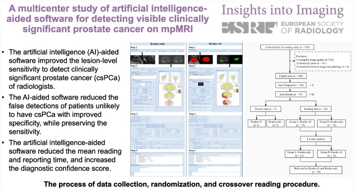
AI-aided software for detecting visible clinically significant prostate cancer on mpMRI

This study seeks to determine if artificial intelligence (AI)-based software can improve radiologists’ performance when detecting clinically significant prostate cancer. Sixteen radiologists from four hospitals participated and were assigned 30 cases, half without AI and half with AI. The authors determined that the AI software improves the performance of radiologists by reducing false positive detection of prostate cancer patients while

Read More →

Machine learning–based radiomics classifies parotid tumors using morphological MRI

This comparative study aimed to evaluate the effectiveness of machine learning models based on morphological MRI radiomics in the classification of parotid tumors. The authors developed three-step machine learning models with extreme gradient boosting (XGBoost), support vector machine (SVM), and decision tree (DT) algorithms in order to classify the parotid neoplasms into four subtypes. The study was able to demonstrate

Read More →

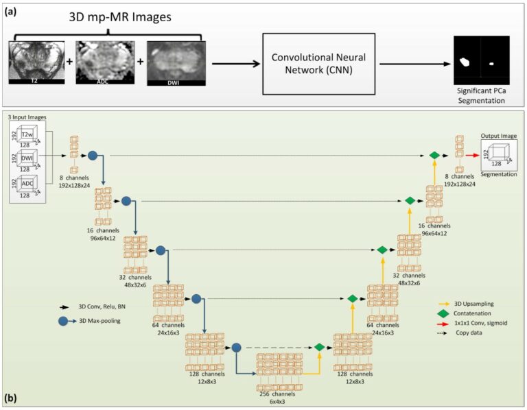
Clinically significant prostate cancer detection and segmentation in low-risk patients using a convolutional neural network on multi-parametric MRI

The purpose of this study was to develop an automatic method for the identification and segmentation of clinically significant prostate cancer in low-risk patients and evaluate this performance in a routine clinical setting. The authors discovered that the proposed deep learning computer-aided method showed promising results in the previously-mentioned identification and segmentation of clinically significant prostate cancer in patients on

Read More →

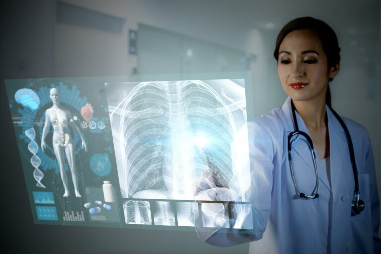
AI in the clinical field of lung disease and COVID-19

Artificial intelligence (AI) tools are becoming a common occurrence in everyday healthcare, especially in the areas of early detection and diagnosis. As lung diseases and the current COVID-19 pandemic become more prevalent around the world, early detection and diagnosis can be literal lifesavers. AI, along with collaborative efforts of the radiological, healthcare industry, and academic communities, are helping to make

Read More →

Can training data help radiologists to open deep learning black box?

Deep learning has recently pervaded the radiology field, reaching promising results that have encouraged both scientists and entrepreneurs to apply these models to improve patient care. However, “with great power there must also come — great responsibility” [1]! In most cases, the complexity of deep learning models forces their users, and sometimes also their developers, to treat them as black

Read More →

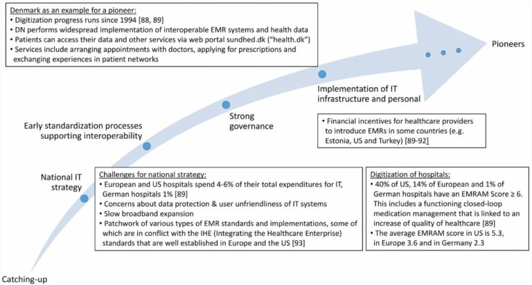
Implementation of eHealth and AI integrated diagnostics with multidisciplinary digitized data: are we ready from an international perspective?

Increasing numbers of publications document the potential of radiomics and artificial intelligence (AI)-aided diagnostics, and the Corona pandemic made the importance of digitally well-connected health systems even clearer. However, although the essential technologies are already available, the progress into clinical routine application is considerably slow. Thus, we intended to perform a comprehensive analysis to learn from the experiences of pioneering

Read More →

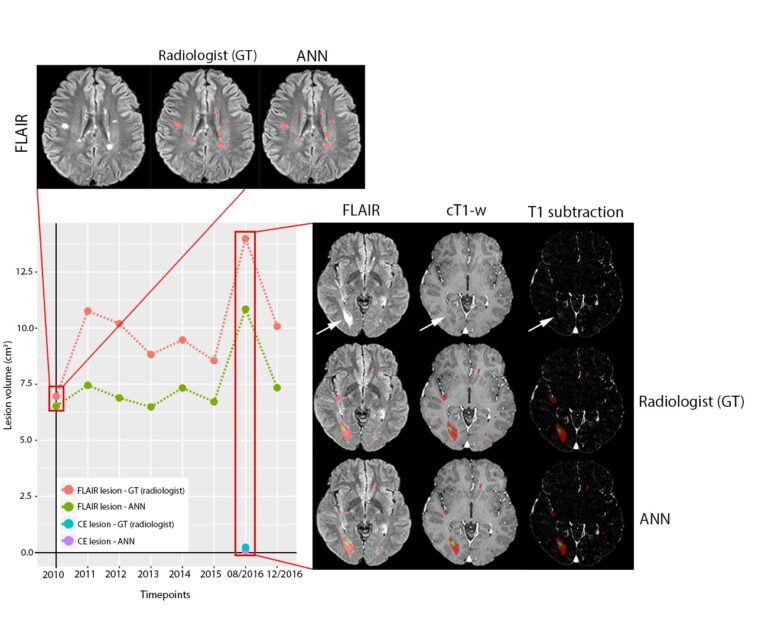
Automated volumetric assessment with artificial neural networks might enable a more accurate assessment of disease burden in patients with multiple sclerosis

Multiple automated methods for segmentation of multiple sclerosis (MS) lesions have been developed over the past years, and the use of artificial neural networks (ANN) has recently generated many outstanding results in the public segmentation challenges. As we all know from our work as radiologists, the routine clinical practice is always conducted with an economical balance between optimal scan times

Read More →

Radiomics analysis using contrast-enhanced CT for preoperative prediction of occult peritoneal metastasis in advanced gastric cancer

The authors of this study constructed two multivariate logistic regression models and compared the diagnostic performance between the two of them via receiver operating characteristic analysis, discovering that CT radiomics analysis can provide valuable information for predicting occult peritoneal metastases in advanced gastric cancer. Key points Venous CT radiomics analysis provided valuable information for predicting occult peritoneal metastases in advanced

Read More →

AI-aided software for detecting visible clinically significant prostate cancer on mpMRI

This study seeks to determine if artificial intelligence (AI)-based software can improve radiologists’ performance when detecting clinically significant prostate cancer. Sixteen radiologists from four hospitals participated and were assigned 30 cases, half without AI and half with AI. The authors determined that the AI software improves the performance of radiologists by reducing false positive detection of prostate cancer patients while

Read More →

Machine learning–based radiomics classifies parotid tumors using morphological MRI

This comparative study aimed to evaluate the effectiveness of machine learning models based on morphological MRI radiomics in the classification of parotid tumors. The authors developed three-step machine learning models with extreme gradient boosting (XGBoost), support vector machine (SVM), and decision tree (DT) algorithms in order to classify the parotid neoplasms into four subtypes. The study was able to demonstrate

Read More →

Clinically significant prostate cancer detection and segmentation in low-risk patients using a convolutional neural network on multi-parametric MRI

The purpose of this study was to develop an automatic method for the identification and segmentation of clinically significant prostate cancer in low-risk patients and evaluate this performance in a routine clinical setting. The authors discovered that the proposed deep learning computer-aided method showed promising results in the previously-mentioned identification and segmentation of clinically significant prostate cancer in patients on

Read More →

AI in the clinical field of lung disease and COVID-19

Artificial intelligence (AI) tools are becoming a common occurrence in everyday healthcare, especially in the areas of early detection and diagnosis. As lung diseases and the current COVID-19 pandemic become more prevalent around the world, early detection and diagnosis can be literal lifesavers. AI, along with collaborative efforts of the radiological, healthcare industry, and academic communities, are helping to make

Read More →

Can training data help radiologists to open deep learning black box?

Deep learning has recently pervaded the radiology field, reaching promising results that have encouraged both scientists and entrepreneurs to apply these models to improve patient care. However, “with great power there must also come — great responsibility” [1]! In most cases, the complexity of deep learning models forces their users, and sometimes also their developers, to treat them as black

Read More →

Implementation of eHealth and AI integrated diagnostics with multidisciplinary digitized data: are we ready from an international perspective?

Increasing numbers of publications document the potential of radiomics and artificial intelligence (AI)-aided diagnostics, and the Corona pandemic made the importance of digitally well-connected health systems even clearer. However, although the essential technologies are already available, the progress into clinical routine application is considerably slow. Thus, we intended to perform a comprehensive analysis to learn from the experiences of pioneering

Read More →

Automated volumetric assessment with artificial neural networks might enable a more accurate assessment of disease burden in patients with multiple sclerosis

Multiple automated methods for segmentation of multiple sclerosis (MS) lesions have been developed over the past years, and the use of artificial neural networks (ANN) has recently generated many outstanding results in the public segmentation challenges. As we all know from our work as radiologists, the routine clinical practice is always conducted with an economical balance between optimal scan times

Read More →

Radiomics analysis using contrast-enhanced CT for preoperative prediction of occult peritoneal metastasis in advanced gastric cancer

The authors of this study constructed two multivariate logistic regression models and compared the diagnostic performance between the two of them via receiver operating characteristic analysis, discovering that CT radiomics analysis can provide valuable information for predicting occult peritoneal metastases in advanced gastric cancer. Key points Venous CT radiomics analysis provided valuable information for predicting occult peritoneal metastases in advanced

Read More →

Become A Member Today!

You will have access to a wide range of benefits that can help you advance your career and stay up-to-date with the latest developments in the field of radiology. These benefits include access to educational resources, networking opportunities with other professionals in the field, opportunities to participate in research projects and clinical trials, and access to the latest technologies and techniques. 

Check out our different membership options.

If you don’t find a fitting membership send us an email here.

Membership

for radiologists, radiology residents, professionals of allied sciences (including radiographers/radiological technologists, nuclear medicine physicians, medical physicists, and data scientists) & professionals of allied sciences in training residing within the boundaries of Europe

  • Reduced registration fees for ECR 1
  • Reduced fees for the European School of Radiology (ESOR) 2
  • Exclusive option to participate in the European Diploma. 3
  • Free electronic access to the journal European Radiology 4
  • Content e-mails for all ESR journals
  • Updates on offers & events through our newsletters
  • Exclusive access to the ESR feed in Juisci

€ 11 /year

Yes! That is less than €1 per month.

Free membership

for radiologists, radiology residents or professionals of allied sciences engaged in practice, teaching or research residing outside Europe as well as individual qualified professionals with an interest in radiology and medical imaging who do not fulfil individual or all requirements for any other ESR membership category & former full members who have retired from all clinical practice
  • Reduced registration fees for ECR 1
  • Free electronic access to the journal European Radiology
  • Content e-mails for all 3 ESR journals 4
  • Updates on offers & events through our newsletters
  • Exclusive access to the ESR feed in Juisci

€ 0

The best things in life are free.

ESR Friends

For students, company representatives or hospital managers etc.

  • Content e-mails for all 3 ESR journals 4
  • Updates on offers & events through our newsletters

€ 0

Friendship doesn’t cost a thing.

The membership type best fitting for you will be selected automatically during the application process.

Footnotes:

01

Reduced registration fees for ECR 2024:
Provided that ESR 2023 membership is activated and approved by August 31, 2023.

Reduced registration fees for ECR 2025:
Provided that ESR 2024 membership is activated and approved by August 31, 2024.

02
Not all activities included
03
Examination based on the ESR European Training Curriculum (radiologists or radiology residents).
04
European Radiology, Insights into Imaging, European Radiology Experimental.