machine learning

Welcome to the blog on Artificial Intelligence of
the European Society of Radiology

This blog aims at bringing educational and critical perspectives on AI to readers. It should help imaging professionals to learn and keep up to date with the technologies being developed in this rapidly evolving field.

Most used hashtags:

Latest posts

Machine learning classifiers vs. Experienced radiologists: Predicting Gleason pattern 4 prostate cancer

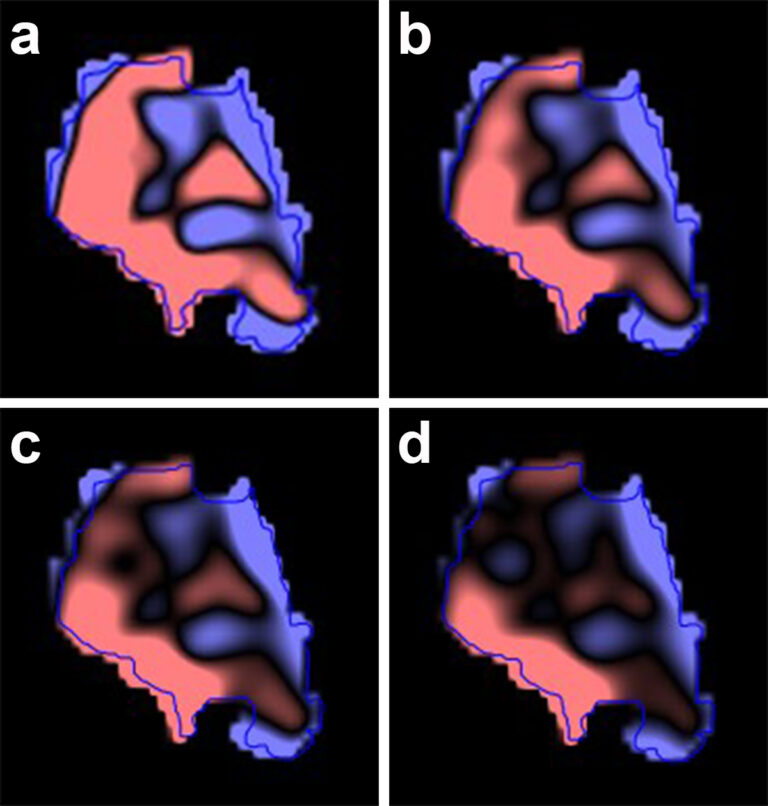
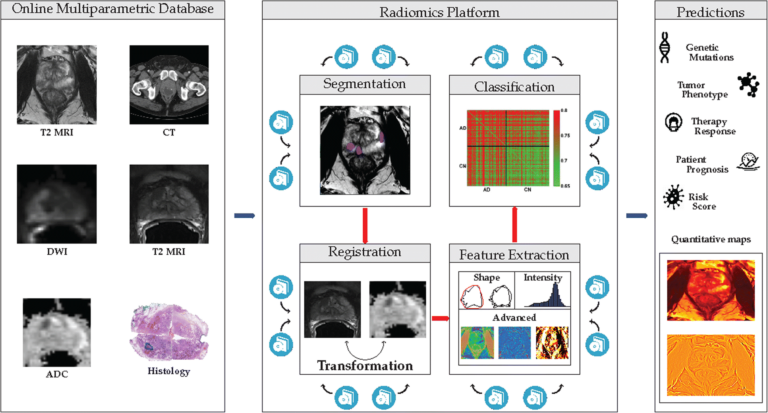
The authors of this study recognized the potential of multiparametric positron emission tomography/magnetic resonance imaging (mpPET/MRI) for detecting and classifying breast lesions. To better understand computer-aided segmentation and diagnosis (CAD) features, the authors introduced a data-driven machine learning approach for a CAD system that enables the assessment of the relevance of mpPET/MRI features on segmentation and classification accuracy. Key points:

Read More →

Data control with AI: who’s in charge?

With the increased use of artificial intelligence (AI) in every segment of life including healthcare, patient data is being collected massively and shared by different stakeholders. But who has control over it? The hospital, the patient, the equipment provider, the software developer, the state’s authorities? If no one knows who controls the data, what can happen? Peter van Ooijen, a

Read More →

Automatic segmentation and classification of breast lesions through identification of informative multiparametric PET/MRI features

The authors of this study recognized the potential of multiparametric positron emission tomography/magnetic resonance imaging (mpPET/MRI) for detecting and classifying breast lesions. To better understand computer-aided segmentation and diagnosis (CAD) features, the authors introduced a data-driven machine learning approach for a CAD system that enables the assessment of the relevance of mpPET/MRI features on segmentation and classification accuracy. Key points:

Read More →

AI guru gives a glimpse into the future of radiology with AI

With the advent of Artificial Intelligence, new opportunities will arise for radiologists if they remain focused and critical, ‘rock star of the digital revolution’ Toby Walsh told delegates at the ESR AI Premium event. A new continent Artificial intelligence (AI) is going to transform every aspect of human life and could generate 15.7 trillion USD – almost China and India’s

Read More →

Application of a deep learning algorithm for detection and visualization of hip fractures on plain pelvic radiographs

In this study, we developed the DCNN not only for the automated detection of hip fractures on frontal pelvic radiographs but also to offer visualization of the fracture site by Grad-CAM, which enables the rapid integration of this tool into the current medical system. The age of artificial intelligence (AI) offers new opportunities but also poses challenges, for physicians. Deep

Read More →

Deep learning reconstruction improves image quality of abdominal ultra-high-resolution CT

Deep learning reconstruction (DLR) is a novel method of reconstruction that introduces deep convolutional neural networks into the reconstruction flow. The authors of this research examined the clinical applicability of abdominal ultra-high-resolution CT (U-HRCT) exams reconstructed with a new DLR and to compare it to hybrid and model-based iterative reconstruction (hybrid-IR, MBIR). Radiologists analysed and graded results from 46 patients

Read More →

Using machine learning to predict cervical lymph node metastasis on dual-energy CT

In this article, we extracted “hand-crafted” radiomic features from dual-energy CT (DECT) virtual monochromatic images (VMIs) reconstructed at different energies and used machine learning to construct prediction models that use the radiomic features of head and neck squamous cell carcinoma (HNSCC) to predict associated nodal metastases. This proof of concept study demonstrated that (1) HNSCC radiomic features can predict associated

Read More →

How machine learning-based high-dimensional CT texture analysis is influenced by segmentation margin

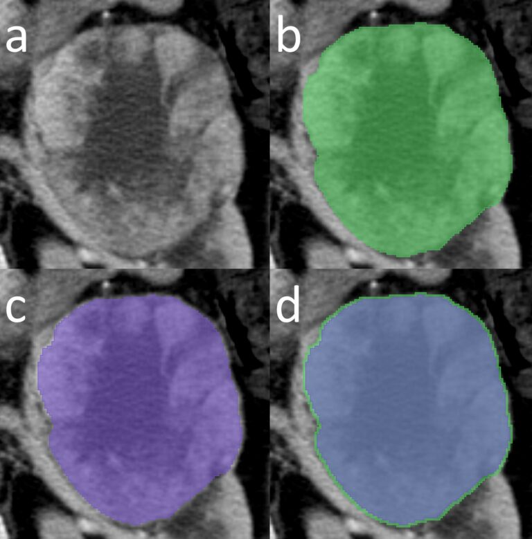
Radiomic workflows include various challenging steps. One of the most demanding steps in radiomics is the segmentation process. Particularly for the renal cell carcinomas, most of the studies used manual tumour contour delineation. In this work, our group wanted to perform an experiment by changing the segmentation margin a little bit, that is, just 2 mm, to see what happens

Read More →

Strategic research agenda for biomedical imaging

Over the last years, medicine has been moving further towards providing a more tailored, patient-centric approach by taking into account as much information as possible to deliver personalised solutions for the individual patient; certainly, radiology and other imaging-based technologies have facilitated this to a great extent. But what will be the way for the future? What are the challenges that

Read More →

Applying 3D CNN to CTA source images to detect ischemic stroke

In this study, the authors investigated how feasible it was to use 3D convolutional neural networks (CNN) to detect ischemic stroke from computed tomography angiography source images (CTA-SI). The study used CTA-SI from 60 randomly selected patients who had a suspected acute ischemic stroke of the middle cerebral artery; half of the patients were used in the neural network training,

Read More →

Machine learning classifiers vs. Experienced radiologists: Predicting Gleason pattern 4 prostate cancer

The authors of this study recognized the potential of multiparametric positron emission tomography/magnetic resonance imaging (mpPET/MRI) for detecting and classifying breast lesions. To better understand computer-aided segmentation and diagnosis (CAD) features, the authors introduced a data-driven machine learning approach for a CAD system that enables the assessment of the relevance of mpPET/MRI features on segmentation and classification accuracy. Key points:

Read More →

Data control with AI: who’s in charge?

With the increased use of artificial intelligence (AI) in every segment of life including healthcare, patient data is being collected massively and shared by different stakeholders. But who has control over it? The hospital, the patient, the equipment provider, the software developer, the state’s authorities? If no one knows who controls the data, what can happen? Peter van Ooijen, a

Read More →

Automatic segmentation and classification of breast lesions through identification of informative multiparametric PET/MRI features

The authors of this study recognized the potential of multiparametric positron emission tomography/magnetic resonance imaging (mpPET/MRI) for detecting and classifying breast lesions. To better understand computer-aided segmentation and diagnosis (CAD) features, the authors introduced a data-driven machine learning approach for a CAD system that enables the assessment of the relevance of mpPET/MRI features on segmentation and classification accuracy. Key points:

Read More →

AI guru gives a glimpse into the future of radiology with AI

With the advent of Artificial Intelligence, new opportunities will arise for radiologists if they remain focused and critical, ‘rock star of the digital revolution’ Toby Walsh told delegates at the ESR AI Premium event. A new continent Artificial intelligence (AI) is going to transform every aspect of human life and could generate 15.7 trillion USD – almost China and India’s

Read More →

Application of a deep learning algorithm for detection and visualization of hip fractures on plain pelvic radiographs

In this study, we developed the DCNN not only for the automated detection of hip fractures on frontal pelvic radiographs but also to offer visualization of the fracture site by Grad-CAM, which enables the rapid integration of this tool into the current medical system. The age of artificial intelligence (AI) offers new opportunities but also poses challenges, for physicians. Deep

Read More →

Deep learning reconstruction improves image quality of abdominal ultra-high-resolution CT

Deep learning reconstruction (DLR) is a novel method of reconstruction that introduces deep convolutional neural networks into the reconstruction flow. The authors of this research examined the clinical applicability of abdominal ultra-high-resolution CT (U-HRCT) exams reconstructed with a new DLR and to compare it to hybrid and model-based iterative reconstruction (hybrid-IR, MBIR). Radiologists analysed and graded results from 46 patients

Read More →

Using machine learning to predict cervical lymph node metastasis on dual-energy CT

In this article, we extracted “hand-crafted” radiomic features from dual-energy CT (DECT) virtual monochromatic images (VMIs) reconstructed at different energies and used machine learning to construct prediction models that use the radiomic features of head and neck squamous cell carcinoma (HNSCC) to predict associated nodal metastases. This proof of concept study demonstrated that (1) HNSCC radiomic features can predict associated

Read More →

How machine learning-based high-dimensional CT texture analysis is influenced by segmentation margin

Radiomic workflows include various challenging steps. One of the most demanding steps in radiomics is the segmentation process. Particularly for the renal cell carcinomas, most of the studies used manual tumour contour delineation. In this work, our group wanted to perform an experiment by changing the segmentation margin a little bit, that is, just 2 mm, to see what happens

Read More →

Strategic research agenda for biomedical imaging

Over the last years, medicine has been moving further towards providing a more tailored, patient-centric approach by taking into account as much information as possible to deliver personalised solutions for the individual patient; certainly, radiology and other imaging-based technologies have facilitated this to a great extent. But what will be the way for the future? What are the challenges that

Read More →

Applying 3D CNN to CTA source images to detect ischemic stroke

In this study, the authors investigated how feasible it was to use 3D convolutional neural networks (CNN) to detect ischemic stroke from computed tomography angiography source images (CTA-SI). The study used CTA-SI from 60 randomly selected patients who had a suspected acute ischemic stroke of the middle cerebral artery; half of the patients were used in the neural network training,

Read More →

Become A Member Today!

You will have access to a wide range of benefits that can help you advance your career and stay up-to-date with the latest developments in the field of radiology. These benefits include access to educational resources, networking opportunities with other professionals in the field, opportunities to participate in research projects and clinical trials, and access to the latest technologies and techniques. 

Check out our different membership options.

If you don’t find a fitting membership send us an email here.

Membership

for radiologists, radiology residents, professionals of allied sciences (including radiographers/radiological technologists, nuclear medicine physicians, medical physicists, and data scientists) & professionals of allied sciences in training residing within the boundaries of Europe

  • Reduced registration fees for ECR 1
  • Reduced fees for the European School of Radiology (ESOR) 2
  • Exclusive option to participate in the European Diploma. 3
  • Free electronic access to the journal European Radiology 4
  • Content e-mails for all ESR journals
  • Updates on offers & events through our newsletters
  • Exclusive access to the ESR feed in Juisci

€ 11 /year

Yes! That is less than €1 per month.

Free membership

for radiologists, radiology residents or professionals of allied sciences engaged in practice, teaching or research residing outside Europe as well as individual qualified professionals with an interest in radiology and medical imaging who do not fulfil individual or all requirements for any other ESR membership category & former full members who have retired from all clinical practice
  • Reduced registration fees for ECR 1
  • Free electronic access to the journal European Radiology
  • Content e-mails for all 3 ESR journals 4
  • Updates on offers & events through our newsletters
  • Exclusive access to the ESR feed in Juisci

€ 0

The best things in life are free.

ESR Friends

For students, company representatives or hospital managers etc.

  • Content e-mails for all 3 ESR journals 4
  • Updates on offers & events through our newsletters

€ 0

Friendship doesn’t cost a thing.

The membership type best fitting for you will be selected automatically during the application process.

Footnotes:

01

Reduced registration fees for ECR 2024:
Provided that ESR 2023 membership is activated and approved by August 31, 2023.

Reduced registration fees for ECR 2025:
Provided that ESR 2024 membership is activated and approved by August 31, 2024.

02
Not all activities included
03
Examination based on the ESR European Training Curriculum (radiologists or radiology residents).
04
European Radiology, Insights into Imaging, European Radiology Experimental.