mri

Welcome to the blog on Artificial Intelligence of
the European Society of Radiology

This blog aims at bringing educational and critical perspectives on AI to readers. It should help imaging professionals to learn and keep up to date with the technologies being developed in this rapidly evolving field.

Most used hashtags:

Latest posts

T2-weighted MRI-based radiomics discriminati between benign and borderline epithelial ovarian tumors

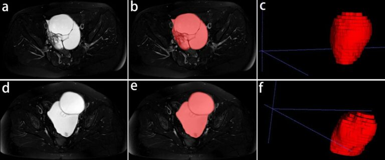
The authors of this study investigated whether radiomics based on T2-weighted MRI was able to discriminate between benign and borderline epithelial ovarian tumors (EOTs) preoperatively. They were able to show that it can provide critical diagnostic information in the discrimination between benign and borderline EOTs, showing that there is potential to aid personalized treatment options. Key points T2-weighted MRI-based radiomics

Read More →

Quality assurance for automatically generated contours with additional deep learning

This study explores the importance of quality assurance when deploying an automatic segmentation model. The authors of this study built a deep-learning model with the goal of estimating the quality of automatically generated contours. They found that the trained model can be used alongside automatic segmentation tools, thus ensuring quality and allowing intervention to prevent undesired segmentation behavior. Key points

Read More →

Application of deep learning to improve image quality and reduce scan time

In this study, the image quality and diagnostic performance of conventional motion-corrected periodically rotated overlapping parallel line with enhanced reconstruction (PROPELLER) MRI sequences was compared with post-processed PROPELLER MRI sequences using deep learning (DL)-based reconstructions. The authors found that the accelerated PROPELLER sequences with DL post-processing showed superior image quality and higher diagnostic confidence when compared to conventional PROPELLER sequences.

Read More →

Tasks for AI in prostate MRI

The authors of this narrative review aimed to introduce quality metrics for emerging artificial intelligence (AI) papers, such as the Checklist for Artificial Intelligence in Medical Imaging (CLAIM) and Field-Weighted Citation Impact (FWCI). Furthermore, the study dives into some of the top AI models for segmentation, detection, and classification, while concluding that prospective studies with multi-center design will need to

Read More →

Prediction of lipomatous soft tissue malignancy on MRI

This study, which included a cohort of 145 patients affected by lipomatous soft tissue tumours, aimed to compare the performances of MRI radiomic machine learning analysis with that of deep learning in order to predict malignancy in patients with lipomas oratypical lipomatous tumours. The authors were able to show that batch-effect corrected machine learning and radiomics approaches outperformed deep learning-based

Read More →

Machine learning–based radiomics classifies parotid tumors using morphological MRI

This comparative study aimed to evaluate the effectiveness of machine learning models based on morphological MRI radiomics in the classification of parotid tumors. The authors developed three-step machine learning models with extreme gradient boosting (XGBoost), support vector machine (SVM), and decision tree (DT) algorithms in order to classify the parotid neoplasms into four subtypes. The study was able to demonstrate

Read More →

The role of generative adversarial networks in brain MRI

Magnetic Resonance Imaging (MRI) is a widely used medical imaging technology that is non-intrusive and considered safe for humans and can generate different modalities of an image, as well as provide valuable insights into a specific disease. The frequent sequences of MRI are T1-weighted and T2- weighted scans. The popularity of artificial intelligence (AI) for brain MRI is on the

Read More →

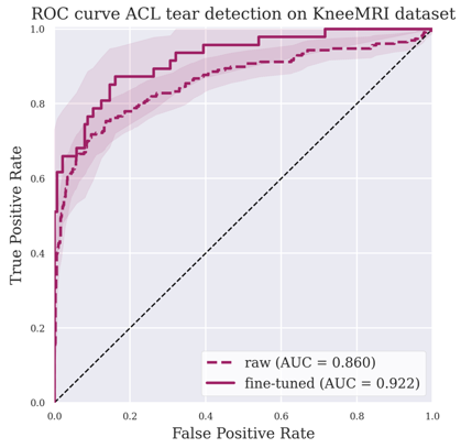
Deep learning detection of ACL tear on knee MRI

The purpose of this study was to develop a deep-learning algorithm for tear detection in the anterior cruciate ligament (ACL), subsequently comparing its accuracy using two external datasets. The authors were able to conclude that their algorithm was capable of showing high performance in the detection of ACL tears. Key points An algorithm for detecting anterior cruciate ligament ruptures was

Read More →

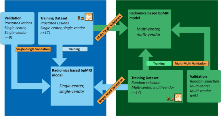
Single-center versus multi-center biparametric MRI radiomics approach

In this study, the authors’ aim was to investigate a previously developed radiomics-based biparametric magnetic resonance imaging (bpMRI) approach used for the discrimination of clinically significant peripheral zone prostate cancer (PZ csPCa) through the use of multi-center, multi-vendor (McMv) and single-center, single-vendor (ScSv) datasets. Using these datasets, the authors were able to determine that a single-center trained radiomics-based bpMRI model

Read More →

Radiomics in the prediction of disease-free survival in early-stage squamous cervical cancer

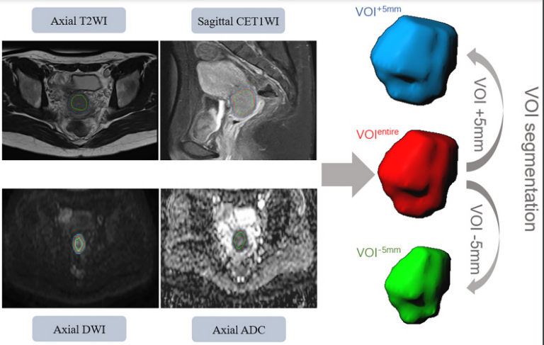
The authors of this study conducted multiparametric magnetic resonance imaging (MRI)-derived radiomics based on multi-scale tumor region in order to predict disease-free survival (DFS) in a cohort of 191 patients with early-stage squamous cervical cancer (ESSCC). They were able to conclude that multiparametric MRI-derived radiomics based on multi-scale tumor region can in fact aid in the prediction of DFS for

Read More →

T2-weighted MRI-based radiomics discriminati between benign and borderline epithelial ovarian tumors

The authors of this study investigated whether radiomics based on T2-weighted MRI was able to discriminate between benign and borderline epithelial ovarian tumors (EOTs) preoperatively. They were able to show that it can provide critical diagnostic information in the discrimination between benign and borderline EOTs, showing that there is potential to aid personalized treatment options. Key points T2-weighted MRI-based radiomics

Read More →

Quality assurance for automatically generated contours with additional deep learning

This study explores the importance of quality assurance when deploying an automatic segmentation model. The authors of this study built a deep-learning model with the goal of estimating the quality of automatically generated contours. They found that the trained model can be used alongside automatic segmentation tools, thus ensuring quality and allowing intervention to prevent undesired segmentation behavior. Key points

Read More →

Application of deep learning to improve image quality and reduce scan time

In this study, the image quality and diagnostic performance of conventional motion-corrected periodically rotated overlapping parallel line with enhanced reconstruction (PROPELLER) MRI sequences was compared with post-processed PROPELLER MRI sequences using deep learning (DL)-based reconstructions. The authors found that the accelerated PROPELLER sequences with DL post-processing showed superior image quality and higher diagnostic confidence when compared to conventional PROPELLER sequences.

Read More →

Tasks for AI in prostate MRI

The authors of this narrative review aimed to introduce quality metrics for emerging artificial intelligence (AI) papers, such as the Checklist for Artificial Intelligence in Medical Imaging (CLAIM) and Field-Weighted Citation Impact (FWCI). Furthermore, the study dives into some of the top AI models for segmentation, detection, and classification, while concluding that prospective studies with multi-center design will need to

Read More →

Prediction of lipomatous soft tissue malignancy on MRI

This study, which included a cohort of 145 patients affected by lipomatous soft tissue tumours, aimed to compare the performances of MRI radiomic machine learning analysis with that of deep learning in order to predict malignancy in patients with lipomas oratypical lipomatous tumours. The authors were able to show that batch-effect corrected machine learning and radiomics approaches outperformed deep learning-based

Read More →

Machine learning–based radiomics classifies parotid tumors using morphological MRI

This comparative study aimed to evaluate the effectiveness of machine learning models based on morphological MRI radiomics in the classification of parotid tumors. The authors developed three-step machine learning models with extreme gradient boosting (XGBoost), support vector machine (SVM), and decision tree (DT) algorithms in order to classify the parotid neoplasms into four subtypes. The study was able to demonstrate

Read More →

The role of generative adversarial networks in brain MRI

Magnetic Resonance Imaging (MRI) is a widely used medical imaging technology that is non-intrusive and considered safe for humans and can generate different modalities of an image, as well as provide valuable insights into a specific disease. The frequent sequences of MRI are T1-weighted and T2- weighted scans. The popularity of artificial intelligence (AI) for brain MRI is on the

Read More →

Deep learning detection of ACL tear on knee MRI

The purpose of this study was to develop a deep-learning algorithm for tear detection in the anterior cruciate ligament (ACL), subsequently comparing its accuracy using two external datasets. The authors were able to conclude that their algorithm was capable of showing high performance in the detection of ACL tears. Key points An algorithm for detecting anterior cruciate ligament ruptures was

Read More →

Single-center versus multi-center biparametric MRI radiomics approach

In this study, the authors’ aim was to investigate a previously developed radiomics-based biparametric magnetic resonance imaging (bpMRI) approach used for the discrimination of clinically significant peripheral zone prostate cancer (PZ csPCa) through the use of multi-center, multi-vendor (McMv) and single-center, single-vendor (ScSv) datasets. Using these datasets, the authors were able to determine that a single-center trained radiomics-based bpMRI model

Read More →

Radiomics in the prediction of disease-free survival in early-stage squamous cervical cancer

The authors of this study conducted multiparametric magnetic resonance imaging (MRI)-derived radiomics based on multi-scale tumor region in order to predict disease-free survival (DFS) in a cohort of 191 patients with early-stage squamous cervical cancer (ESSCC). They were able to conclude that multiparametric MRI-derived radiomics based on multi-scale tumor region can in fact aid in the prediction of DFS for

Read More →

Become A Member Today!

You will have access to a wide range of benefits that can help you advance your career and stay up-to-date with the latest developments in the field of radiology. These benefits include access to educational resources, networking opportunities with other professionals in the field, opportunities to participate in research projects and clinical trials, and access to the latest technologies and techniques. 

Check out our different membership options.

If you don’t find a fitting membership send us an email here.

Membership

for radiologists, radiology residents, professionals of allied sciences (including radiographers/radiological technologists, nuclear medicine physicians, medical physicists, and data scientists) & professionals of allied sciences in training residing within the boundaries of Europe

  • Reduced registration fees for ECR 1
  • Reduced fees for the European School of Radiology (ESOR) 2
  • Exclusive option to participate in the European Diploma. 3
  • Free electronic access to the journal European Radiology 4
  • Content e-mails for all ESR journals
  • Updates on offers & events through our newsletters
  • Exclusive access to the ESR feed in Juisci

€ 11 /year

Yes! That is less than €1 per month.

Free membership

for radiologists, radiology residents or professionals of allied sciences engaged in practice, teaching or research residing outside Europe as well as individual qualified professionals with an interest in radiology and medical imaging who do not fulfil individual or all requirements for any other ESR membership category & former full members who have retired from all clinical practice
  • Reduced registration fees for ECR 1
  • Free electronic access to the journal European Radiology
  • Content e-mails for all 3 ESR journals 4
  • Updates on offers & events through our newsletters
  • Exclusive access to the ESR feed in Juisci

€ 0

The best things in life are free.

ESR Friends

For students, company representatives or hospital managers etc.

  • Content e-mails for all 3 ESR journals 4
  • Updates on offers & events through our newsletters

€ 0

Friendship doesn’t cost a thing.

The membership type best fitting for you will be selected automatically during the application process.

Footnotes:

01

Reduced registration fees for ECR 2024:
Provided that ESR 2023 membership is activated and approved by August 31, 2023.

Reduced registration fees for ECR 2025:
Provided that ESR 2024 membership is activated and approved by August 31, 2024.

02
Not all activities included
03
Examination based on the ESR European Training Curriculum (radiologists or radiology residents).
04
European Radiology, Insights into Imaging, European Radiology Experimental.