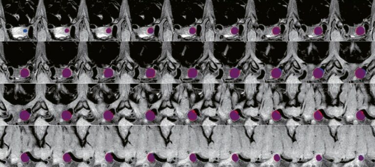
AI Blog

Welcome to the blog on Artificial Intelligence of
the European Society of Radiology

This blog aims at bringing educational and critical perspectives on AI to readers. It should help imaging professionals to learn and keep up to date with the technologies being developed in this rapidly evolving field.

Most used hashtags:

Latest posts

Avoiding Radiology’s Boeing 737 Max moment

Recently, Kemper et al. published an insightful paper on the challenges and opportunities of health technology assessment (HTA) models to evaluate the value, safety, and trustworthiness of radiology computer vision AI (RCVAI) tools used in daily practice. Formal HTA frameworks are theoretically accurate and help define complex issues, but they are ideal approaches. The real world is messier. Ideal descriptions

Read More →

Image biomarkers and explainable AI

Feature extraction and selection in medical data are crucial for radiomics and image biomarker discovery, particularly using convolutional neural networks (CNNs). The process involves feature extraction, dimensionality reduction, and addressing the curse of dimensionality. While deep learning (DL) techniques perform well, handcrafted features are important for certain studies and need to be considered. Dataset size and diversity are also key

Read More →

On Artificial Intelligence: An interview with Daniel Truhn

Following a fellowship in artificial intelligence (AI) at the Institute of Imaging and Computer Vision at RWTH Aachen, which only solidified his belief in AI’s great potential in medicine, Daniel Truhn, a Diagnostic and Interventional radiologist at University Hospital Aachen in Germany, started working on AI’s application in medicine, even creating his own working group in the area. Join us

Read More →

CNNs perform automated staging of cardiac iron overload from multiecho MR sequences

This new European Radiology study aimed to develop a deep-learning model for classifying myocardial iron overload (MIO) using magnitude T2* multi-echo MR images from 496 thalassemia major patients. Two 2D convolutional neural networks (CNN), MS-HippoNet (multi-slice) and SS-HippoNet (single-slice), were trained using 5-fold cross-validation. The model showed strong performance with a multi-class accuracy of 0.885 and 0.836 on test sets,

Read More →

Deep learning in diagnostic imaging of spondyloarthropathies

Our recent systematic review observed that deep learning models for spondyloarthropathies (SpA) hold remarkable promise—not just for identifying subtle radiographic or MRI findings, but also for harnessing massive datasets to detect early, even preclinical, disease features. These techniques can parse millions of pixel-level patterns, potentially flagging nuanced changes that may precede clinical symptoms. Such a capability could fundamentally shift SpA

Read More →

Multi-scene model shows success for precise preoperative and intraoperative segmentation of acute VCFs

This study aimed to develop a multi-scene model, Positioning and Focus Network (PFNet), for automatically segmenting acute vertebral compression fractures (VCFs) from spine radiographs. Conducted across five hospitals from 2016 to 2019, the study included both acute VCF patients and healthy controls. PFNet was trained with radiographs from Hospitals A and B, and tested on datasets from Hospitals A-E. The

Read More →

An AI deep learning algorithm for detecting pulmonary nodules on ultra-low-dose CT in an emergency setting

The OPTIMACT trial investigated the added value of ultra-low-dose CT (ULDCT) compared to chest x-ray in patients suspected of pulmonary disease at the emergency department (ED). As expected, more incidental pulmonary nodules required follow-up in the ULDCT-arm (4.5%) compared to the CXR-arm (0.6%) of the trial. The number of these nodules in the ULDCT-arm was lower than reported in other

Read More →

Using commercial AI-based mammography analysis software to improve breast US interpretations

This study evaluated the use of commercial AI-based mammography software to improve breast ultrasound (US) lesion interpretations. The AI software, which provides an AI malignancy score ranging from 0 to 100, was tested on 1,109 breasts that underwent both mammography and US-guided breast biopsy. The AI software showed an area under the curve (AUROC) of 0.79 for distinguishing benign from

Read More →

Deep learning and compressed sensing for reconstruction of 3D knee MRI

This study explores combining compressed sensing (CS) and artificial intelligence (AI), particularly deep learning (DL), to accelerate three-dimensional (3D) magnetic resonance imaging (MRI) of the knee. Twenty healthy volunteers were examined using a 3-T scanner with a fat-saturated 3D proton density sequence at various acceleration levels. Two reconstruction methods were compared: conventional CS and a new DL-based algorithm (CS-AI). Two

Read More →

On Artificial Intelligence: An interview with Christian Blüthgen

We were delighted to speak with Christian Blüthgen, a radiologist at the Institute for Diagnostic and Interventional Radiology at University Hospital Zurich. Blüthgen was previously a postdoctoral research fellow at the Center for Artificial Intelligence in Medicine and Imaging (AIMI) at Stanford University in California, USA, where he gained practical experience in the design and implementation of deep learning pipelines

Read More →

Become A Member Today!

You will have access to a wide range of benefits that can help you advance your career and stay up-to-date with the latest developments in the field of radiology. These benefits include access to educational resources, networking opportunities with other professionals in the field, opportunities to participate in research projects and clinical trials, and access to the latest technologies and techniques. 

Check out our different membership options.

If you don’t find a fitting membership send us an email here.

Membership

for radiologists, radiology residents, professionals of allied sciences (including radiographers/radiological technologists, nuclear medicine physicians, medical physicists, and data scientists) & professionals of allied sciences in training residing within the boundaries of Europe

  • Reduced registration fees for ECR 1
  • Reduced fees for the European School of Radiology (ESOR) 2
  • Option to participate in the European Diploma. 3
  • Free electronic access to the journal European Radiology 
  • Content e-mails for all ESR journals4
  • Updates on offers & events through our newsletters
  • Exclusive access to the ESR feed in Juisci

€ 11 /year

Yes! That is less than €1 per month.

Free membership

for radiologists, radiology residents or professionals of allied sciences engaged in practice, teaching or research residing outside Europe as well as individual qualified professionals with an interest in radiology and medical imaging who do not fulfil individual or all requirements for any other ESR membership category & former full members who have retired from all clinical practice
  • Reduced registration fees for ECR 1
  • Option to participate in the European Diploma. 3
  • Free electronic access to the journal European Radiology
  • Content e-mails for all ESR journals 4
  • Updates on offers & events through our newsletters
  • Exclusive access to the ESR feed in Juisci

€ 0

The best things in life are free.

ESR Friends

For students, company representatives or hospital managers etc.

  • Content e-mails for all 3 ESR journals 4
  • Updates on offers & events through our newsletters

€ 0

Friendship doesn’t cost a thing.

The membership type best fitting for you will be selected automatically during the application process.

Footnotes:

01

Reduced registration fees for ECR 2026:
Provided that ESR 2025 membership is activated and approved by August 31, 2025.

02
Not all activities included
03
Examination based on the ESR European Training Curriculum (radiologists or radiology residents).
04
European Radiology, Insights into Imaging, European Radiology Experimental.