AI Blog

Welcome to the blog on Artificial Intelligence of
the European Society of Radiology

This blog aims at bringing educational and critical perspectives on AI to readers. It should help imaging professionals to learn and keep up to date with the technologies being developed in this rapidly evolving field.

Most used hashtags:

Latest posts

AI-based algorithm offers reliable Cobb angle measurement on routine MRI for degenerative scoliosis patients

Due to how the severity of degenerative scoliosis is assessed, this retrospective study aimed to develop and evaluate the reliability of a novel automatic method that measured Cobb angles on lumbar MRI in degenerative scoliosis (DS) patients. The authors developed a 3D artificial intelligence algorithm that was trained on 447 lumbar MRI. The study concluded that the AI-based algorithm offered

Read More →

Shallow and deep learning classifiers in medical image analysis

The authors of this review aimed to give educational insight into the most accessible and widely employed classifiers in the field of radiology, distinguish between “shallow” learning algorithms, as well as look into “deep” learning architectures such as convolutional neural networks and vision transformers. This review found that machine learning classifiers offer vital information for the development of clinical decision

Read More →

Deep learning-based 3D cerebrovascular segmentation workflow on bright and black blood sequences MRA

Cerebrovascular diseases are seen as a significant threat to human life and health, and the segmentation of brain blood vessels has become a scientific challenge. Therefore, the authors of this study aimed to develop a fully automated deep learning workflow capable of accurate 3D segmentation of cerebral blood vessels using convolutional neural networks (CNNs) and transformer models. The study, conducted

Read More →

AI applications in musculoskeletal imaging

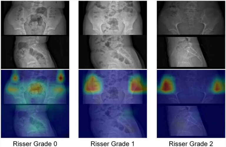
As artificial intelligence (AI) tools and technologies become more ubiquitous in radiology, the role they play in the day-to-day work of radiologists is gaining importance. This narrative review provides an overview of the clinical applications of AI in musculoskeletal imaging, diving into specific areas of musculoskeletal disorders including trauma, bone age estimation, bone and soft-tissue tumors, and orthopedic implant-related pathology.

Read More →

Novel reporting workflow for automated integration of AI results into structured radiology reports

Although artificial intelligence (AI) shows great potential to help radiologists in their daily clinical routine, integrating AI into the radiology workflow is often lacking, underutilizing its full potential. Therefore, this study aimed to develop a new reporting pipeline enabling automated pre-population of structured reports with results provided by commercially available AI tools. The authors successfully demonstrated that the AI to

Read More →

AI in immunotherapy PET/SPECT imaging

Molecular medical imaging, with technologies like PET and SPECT, plays a crucial role in oncology for diagnosis and treatment tracking. Yet, interpreting these images, especially in gauging immunotherapy responses, presents challenges due to new patterns of response and progression as well as inherent subjectivity in image interpretation. While there’s a clear drive to harness artificial intelligence (AI) for improved clinical

Read More →

Sharing is Caring – Promoting Radiomics Research Transparency and Sustainability

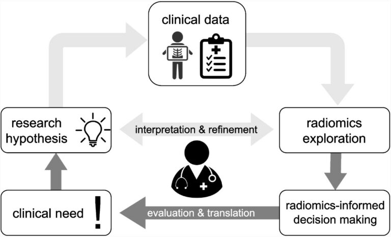
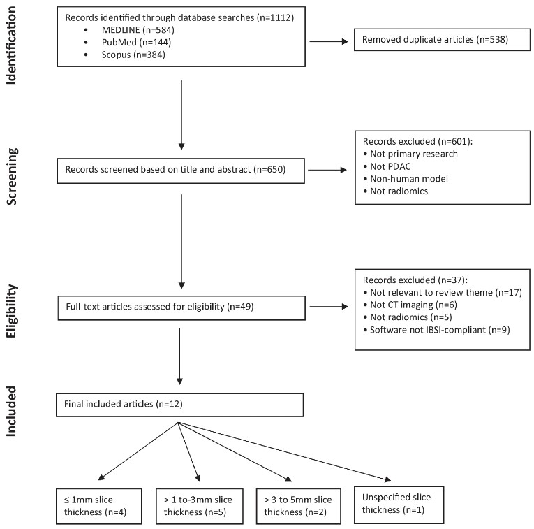
Radiomics, a rapidly growing and innovative field in medical imaging, extracts detailed features from medical scans that could play a pivotal future adjunct role in patient care. However, the clinical adoption of radiomics is stalled by significant research heterogeneity and reproducibility issues [1]. The complex radiomics pipeline, requiring multidisciplinary expertise, often lacks transparency regarding sharing crucial details like software tools

Read More →

Knee landmarks detection via deep learning

A deep learning-based approach was developed and validated in this study which aimed to automatically measure the patellofemoral instability (PFI) indices related to patellar height and trochlear dysplasia in knee MRI scans. The authors included a total of 763 knee MRI slices from 95 patients, annotating 3,393 anatomical landmarks. The results indicated that the developed models achieved good accuracy in

Read More →

Coronary CT angiographic detection of in-stent restenosis via deep learning reconstruction

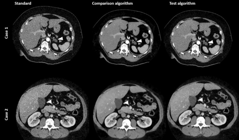
This feasibility study used deep learning reconstruction, Precise IQ Engine (PIQE), to quantify stent strut thickness and lumen vessel diameter, subsequently comparing it with values obtained using conventional reconstruction methods. The authors conducting this study examined 166 stents in 85 consecutive patients who had undergone CT and invasive coronary angiography (ICA) within 3 months of each other. The results demonstrated

Read More →

Deep Learning-based framework shown as a valuable tool to determine the composition of thyroid nodules

The authors of this retrospective multicenter study proposed a deep learning-based framework to identify the composition of thyroid nodules while also assessing their malignancy risk. Their research demonstrated that convolutional neural networks (CNNs) were able to assist in the diagnosis of thyroid nodules and reduce the rate of unnecessary fine-needle aspiration. Key points: Article: Deep learning to assist composition classification

Read More →

Become A Member Today!

You will have access to a wide range of benefits that can help you advance your career and stay up-to-date with the latest developments in the field of radiology. These benefits include access to educational resources, networking opportunities with other professionals in the field, opportunities to participate in research projects and clinical trials, and access to the latest technologies and techniques. 

Check out our different membership options.

If you don’t find a fitting membership send us an email here.

Membership

for radiologists, radiology residents, professionals of allied sciences (including radiographers/radiological technologists, nuclear medicine physicians, medical physicists, and data scientists) & professionals of allied sciences in training residing within the boundaries of Europe

  • Reduced registration fees for ECR 1
  • Reduced fees for the European School of Radiology (ESOR) 2
  • Option to participate in the European Diploma. 3
  • Free electronic access to the journal European Radiology 
  • Content e-mails for all ESR journals4
  • Updates on offers & events through our newsletters
  • Exclusive access to the ESR feed in Juisci

€ 11 /year

Yes! That is less than €1 per month.

Free membership

for radiologists, radiology residents or professionals of allied sciences engaged in practice, teaching or research residing outside Europe as well as individual qualified professionals with an interest in radiology and medical imaging who do not fulfil individual or all requirements for any other ESR membership category & former full members who have retired from all clinical practice
  • Reduced registration fees for ECR 1
  • Option to participate in the European Diploma. 3
  • Free electronic access to the journal European Radiology
  • Content e-mails for all ESR journals 4
  • Updates on offers & events through our newsletters
  • Exclusive access to the ESR feed in Juisci

€ 0

The best things in life are free.

ESR Friends

For students, company representatives or hospital managers etc.

  • Content e-mails for all 3 ESR journals 4
  • Updates on offers & events through our newsletters

€ 0

Friendship doesn’t cost a thing.

The membership type best fitting for you will be selected automatically during the application process.

Footnotes:

01

Reduced registration fees for ECR 2025:
Provided that ESR 2024 membership is activated and approved by August 31, 2024.

02
Not all activities included
03
Examination based on the ESR European Training Curriculum (radiologists or radiology residents).
04
European Radiology, Insights into Imaging, European Radiology Experimental.