deep learning

Welcome to the blog on Artificial Intelligence of
the European Society of Radiology

This blog aims at bringing educational and critical perspectives on AI to readers. It should help imaging professionals to learn and keep up to date with the technologies being developed in this rapidly evolving field.

Most used hashtags:

Latest posts

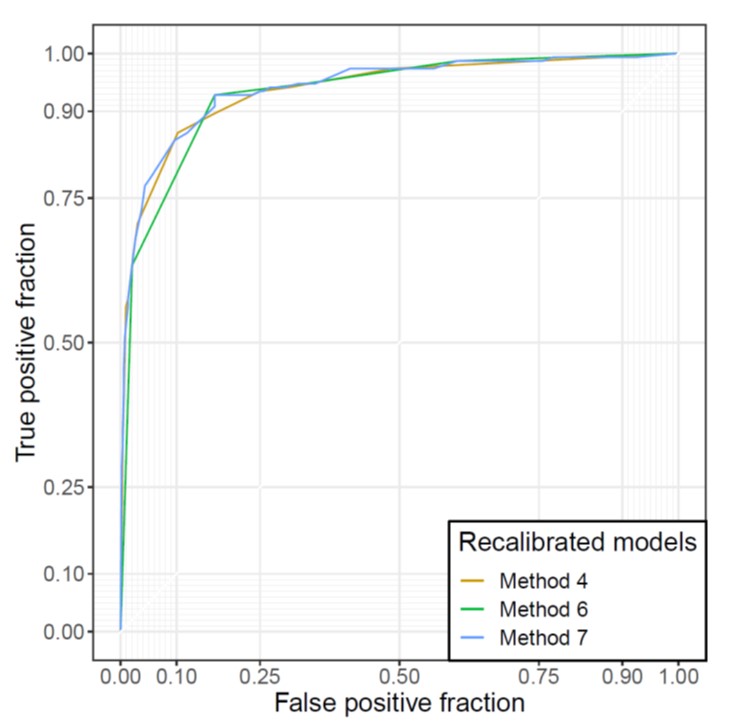
Automated identification of chest radiographs with referable abnormality with deep learning: need for recalibration

The purpose of this study was to evaluate the calibration of a deep learning (DL) model in a diagnostic cohort, as well as to improve the model’s calibration through recalibration procedures. The authors found that the calibration of the DL algorithm can be augmented through simple recalibration procedures, and improved calibration may enhance the interpretability and credibility of the model

Read More →

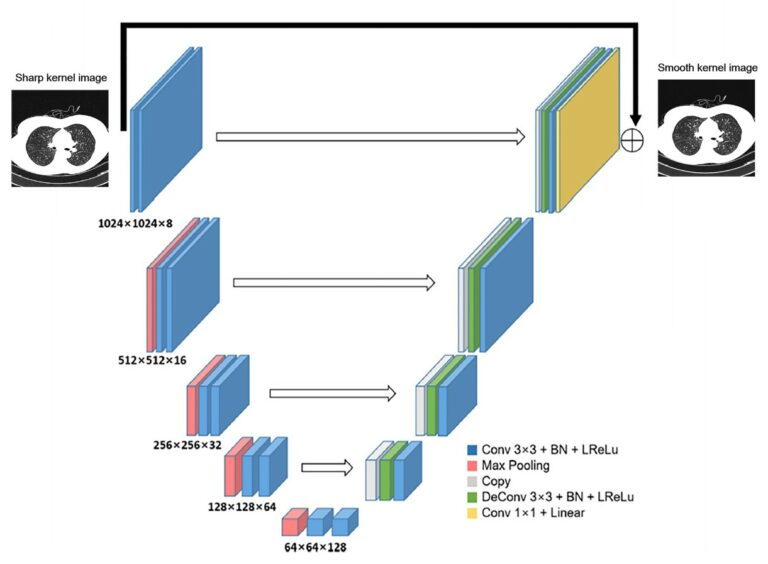
Emphysema quantification using low-dose computed tomography with deep learning-based kernel conversion comparison

This study used a sample of 131 participants who underwent low-dose computed tomography (LDCT) and standard-dose computed tomography (SDCT) to determine the effect of dose reduction and kernel selection on quantifying emphysema. The authors determined that the deep learning-based CT kernel conversation of sharp kernel in LDCT significantly reduced the variation in emphysema quantification. Key points Low-dose computed tomography with

Read More →

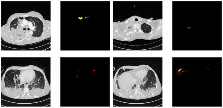
Deep learning detection and quantification of pneumothorax in heterogeneous routine chest computed tomography

In this study, the authors proposed a deep learning method for the detection and quantification of pneumothorax in heterogeneous routine clinical data, which may facilitate the automated triage of urgent examinations and enable support in the treatment decision. Key points Pneumothorax is an important pathology to be included in applications that are designed to triage urgent imaging examinations. Heterogeneity in

Read More →

Deep learning for the determination of myometrial invasion depth and automatic lesion identification in endometrial cancer MR imaging

Endometrial cancer (EC) has the highest rate of malignancy in women in the entire world, including China, which has the largest population. Accurately staging EC prior to an invasive procedure still poses a challenge for clinicians. In the present study, we used more than five hundred EC patients’ MR images to train the computer to establish a deep learning diagnostic

Read More →

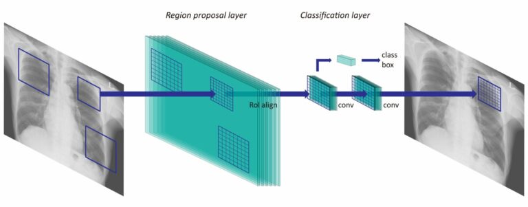
Optimal matrix size of chest radiographs for computer-aided detection on lung nodule or mass with deep learning

In this study, the authors retrospectively collected 2,088 abnormal and 352 normal chest radiographs from two institutions in order to investigate the optimal input matrix size for deep learning-based computer-aided detection (CAD) of nodules and masses on chest radiographs. This resulted in the matrix size 896 as having the highest performance for various sizes of abnormalities using different convolutional neural

Read More →

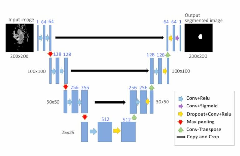
Deep learning for fully automated tumor segmentation and extraction of magnetic resonance radiomics features in cervical cancer

The purpose of this retrospective study was to develop and evaluate the performance of U-Net to determine whether U-Net-based deep learning could accurately perform fully automated localization and segmentation of cervical tumors in MR images, as well as the robustness of extracting apparent diffusion coefficient (ADC) radiomics features. Key points U-Net-based deep learning can perform accurate fully automated localization and

Read More →

Deep learning: definition and perspectives for thoracic imaging

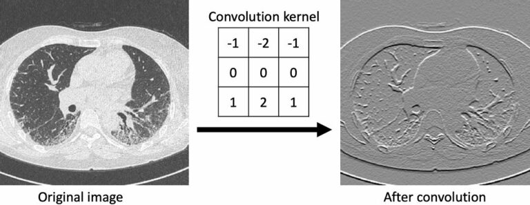
The authors of this review aimed to provide definitions for understanding the methods of machine learning, deep learning, and convolutional neural networks (CNN) and to dive into their roles and potential in the area of thoracic imaging. Key points Deep learning outperforms other machine learning techniques for number of tasks in radiology. Convolutional neural network is the most popular deep

Read More →

Artificial intelligence for detecting small FDG-positive lung nodules in digital PET/CT

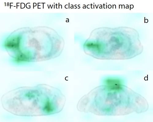
The authors of this study aimed to determine the diagnostic performance of a deep learning algorithm for the automated detection of small 18F-FDG-avid pulmonary nodules in positron emission tomography (PET) scans. Their findings suggest that these machine learning algorithms may be able to aid in the detection of small 18F-FDG-avid pulmonary nodules in clinical PET/CT scans, with artificial intelligence (AI)

Read More →

Accurate prediction of responses to transarterial chemoembolization for patients with hepatocellular carcinoma by using artificial intelligence in contrast-enhanced ultrasound

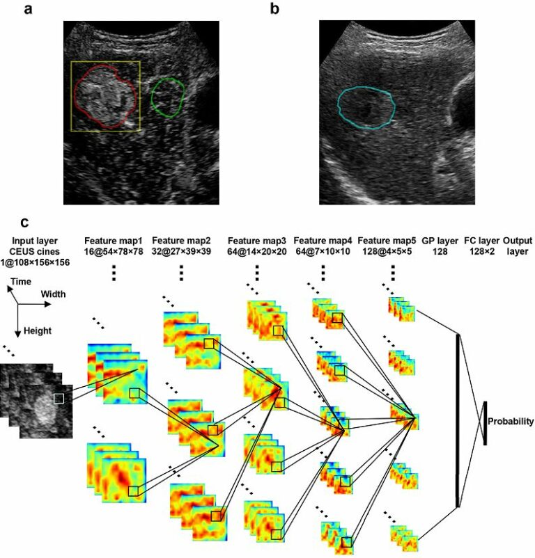
The aim of this study was to establish and validate an artificial intelligence-based radiomics strategy in order to predict personalized responses to hepatocellular carcinoma (HCC) to first transarterial chemoembolization (TACE) by analyzing contrast-enhanced ultrasound (CEUS) cines quantitatively. This was done using 130 HCC patients, showing that a deep learning-based radiomics method can effectively utilize CEUS, resulting in accurate and personalized

Read More →

Deep learning workflow in radiology: how to get started

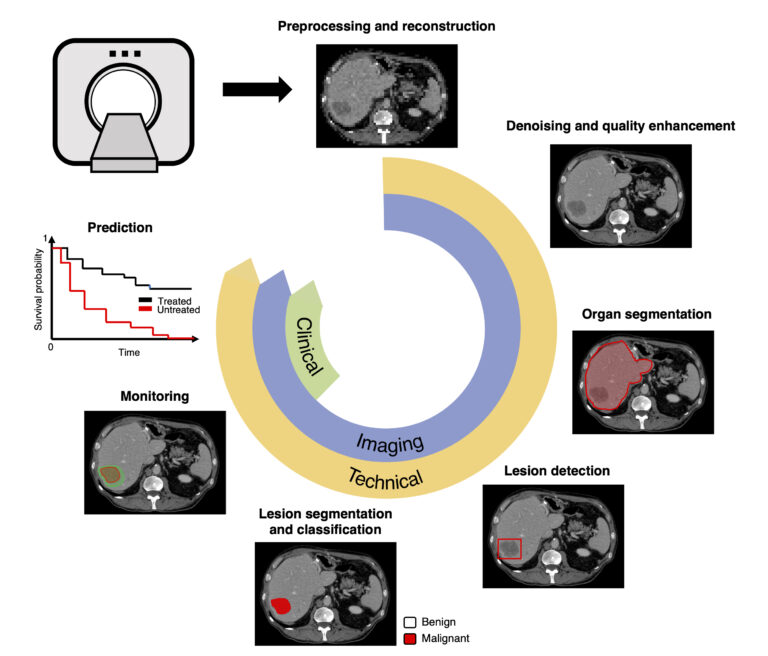
In the past decade, deep learning architectures, which essentially consist of neural networks with numerous layers, have emerged as a dominant class of machine learning algorithms. Owing to the availability of larger datasets in radiology and access to high-performance graphical processing units, deep learning has provided state-of-the-art performance for various computer vision tasks such as lesion detection, segmentation, classification, monitoring,

Read More →

Automated identification of chest radiographs with referable abnormality with deep learning: need for recalibration

The purpose of this study was to evaluate the calibration of a deep learning (DL) model in a diagnostic cohort, as well as to improve the model’s calibration through recalibration procedures. The authors found that the calibration of the DL algorithm can be augmented through simple recalibration procedures, and improved calibration may enhance the interpretability and credibility of the model

Read More →

Emphysema quantification using low-dose computed tomography with deep learning-based kernel conversion comparison

This study used a sample of 131 participants who underwent low-dose computed tomography (LDCT) and standard-dose computed tomography (SDCT) to determine the effect of dose reduction and kernel selection on quantifying emphysema. The authors determined that the deep learning-based CT kernel conversation of sharp kernel in LDCT significantly reduced the variation in emphysema quantification. Key points Low-dose computed tomography with

Read More →

Deep learning detection and quantification of pneumothorax in heterogeneous routine chest computed tomography

In this study, the authors proposed a deep learning method for the detection and quantification of pneumothorax in heterogeneous routine clinical data, which may facilitate the automated triage of urgent examinations and enable support in the treatment decision. Key points Pneumothorax is an important pathology to be included in applications that are designed to triage urgent imaging examinations. Heterogeneity in

Read More →

Deep learning for the determination of myometrial invasion depth and automatic lesion identification in endometrial cancer MR imaging

Endometrial cancer (EC) has the highest rate of malignancy in women in the entire world, including China, which has the largest population. Accurately staging EC prior to an invasive procedure still poses a challenge for clinicians. In the present study, we used more than five hundred EC patients’ MR images to train the computer to establish a deep learning diagnostic

Read More →

Optimal matrix size of chest radiographs for computer-aided detection on lung nodule or mass with deep learning

In this study, the authors retrospectively collected 2,088 abnormal and 352 normal chest radiographs from two institutions in order to investigate the optimal input matrix size for deep learning-based computer-aided detection (CAD) of nodules and masses on chest radiographs. This resulted in the matrix size 896 as having the highest performance for various sizes of abnormalities using different convolutional neural

Read More →

Deep learning for fully automated tumor segmentation and extraction of magnetic resonance radiomics features in cervical cancer

The purpose of this retrospective study was to develop and evaluate the performance of U-Net to determine whether U-Net-based deep learning could accurately perform fully automated localization and segmentation of cervical tumors in MR images, as well as the robustness of extracting apparent diffusion coefficient (ADC) radiomics features. Key points U-Net-based deep learning can perform accurate fully automated localization and

Read More →

Deep learning: definition and perspectives for thoracic imaging

The authors of this review aimed to provide definitions for understanding the methods of machine learning, deep learning, and convolutional neural networks (CNN) and to dive into their roles and potential in the area of thoracic imaging. Key points Deep learning outperforms other machine learning techniques for number of tasks in radiology. Convolutional neural network is the most popular deep

Read More →

Artificial intelligence for detecting small FDG-positive lung nodules in digital PET/CT

The authors of this study aimed to determine the diagnostic performance of a deep learning algorithm for the automated detection of small 18F-FDG-avid pulmonary nodules in positron emission tomography (PET) scans. Their findings suggest that these machine learning algorithms may be able to aid in the detection of small 18F-FDG-avid pulmonary nodules in clinical PET/CT scans, with artificial intelligence (AI)

Read More →

Accurate prediction of responses to transarterial chemoembolization for patients with hepatocellular carcinoma by using artificial intelligence in contrast-enhanced ultrasound

The aim of this study was to establish and validate an artificial intelligence-based radiomics strategy in order to predict personalized responses to hepatocellular carcinoma (HCC) to first transarterial chemoembolization (TACE) by analyzing contrast-enhanced ultrasound (CEUS) cines quantitatively. This was done using 130 HCC patients, showing that a deep learning-based radiomics method can effectively utilize CEUS, resulting in accurate and personalized

Read More →

Deep learning workflow in radiology: how to get started

In the past decade, deep learning architectures, which essentially consist of neural networks with numerous layers, have emerged as a dominant class of machine learning algorithms. Owing to the availability of larger datasets in radiology and access to high-performance graphical processing units, deep learning has provided state-of-the-art performance for various computer vision tasks such as lesion detection, segmentation, classification, monitoring,

Read More →

Become A Member Today!

You will have access to a wide range of benefits that can help you advance your career and stay up-to-date with the latest developments in the field of radiology. These benefits include access to educational resources, networking opportunities with other professionals in the field, opportunities to participate in research projects and clinical trials, and access to the latest technologies and techniques. 

Check out our different membership options.

If you don’t find a fitting membership send us an email here.

Membership

for radiologists, radiology residents, professionals of allied sciences (including radiographers/radiological technologists, nuclear medicine physicians, medical physicists, and data scientists) & professionals of allied sciences in training residing within the boundaries of Europe

  • Reduced registration fees for ECR 1
  • Reduced fees for the European School of Radiology (ESOR) 2
  • Exclusive option to participate in the European Diploma. 3
  • Free electronic access to the journal European Radiology 4
  • Content e-mails for all ESR journals
  • Updates on offers & events through our newsletters
  • Exclusive access to the ESR feed in Juisci

€ 11 /year

Yes! That is less than €1 per month.

Free membership

for radiologists, radiology residents or professionals of allied sciences engaged in practice, teaching or research residing outside Europe as well as individual qualified professionals with an interest in radiology and medical imaging who do not fulfil individual or all requirements for any other ESR membership category & former full members who have retired from all clinical practice
  • Reduced registration fees for ECR 1
  • Free electronic access to the journal European Radiology
  • Content e-mails for all 3 ESR journals 4
  • Updates on offers & events through our newsletters
  • Exclusive access to the ESR feed in Juisci

€ 0

The best things in life are free.

ESR Friends

For students, company representatives or hospital managers etc.

  • Content e-mails for all 3 ESR journals 4
  • Updates on offers & events through our newsletters

€ 0

Friendship doesn’t cost a thing.

The membership type best fitting for you will be selected automatically during the application process.

Footnotes:

01

Reduced registration fees for ECR 2024:
Provided that ESR 2023 membership is activated and approved by August 31, 2023.

Reduced registration fees for ECR 2025:
Provided that ESR 2024 membership is activated and approved by August 31, 2024.

02
Not all activities included
03
Examination based on the ESR European Training Curriculum (radiologists or radiology residents).
04
European Radiology, Insights into Imaging, European Radiology Experimental.