AI Blog

Welcome to the blog on Artificial Intelligence of
the European Society of Radiology

This blog aims at bringing educational and critical perspectives on AI to readers. It should help imaging professionals to learn and keep up to date with the technologies being developed in this rapidly evolving field.

Most used hashtags:

Latest posts

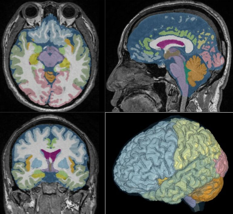
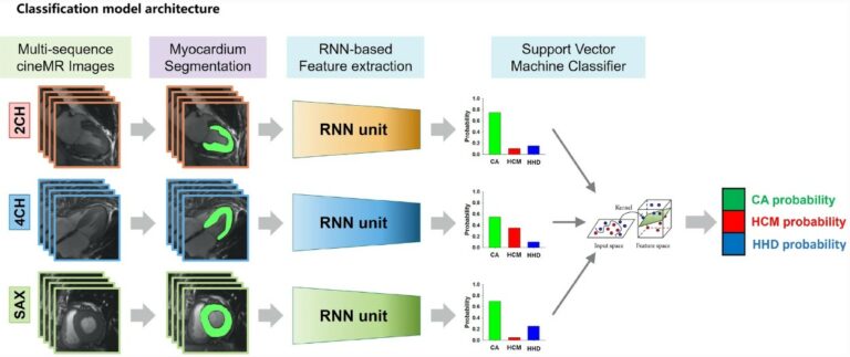
Multi-channel deep learning model diagnoses the cause of LVH

A new study sees the development of a fully automatic framework for the diagnosis of the cause of left ventricular hypertrophy (LVH) via cardiac cine images. The fully automatic myocardium segmentation and spatial-temporal morphology feature-based LVH etiology diagnosis deep learning framework model was able to show a favorable and robust performance in diagnosing the cause of LVH, which could be

Read More →

Evaluating a deep learning software for lung parenchyma characterization in COVID-19 pneumonia

The aim of this study was to evaluate the performance of the LungQuant system, which is a deep learning-based software for quantitative analysis of chest CT. LungQuant was evaluated by comparing its results with independent visual evaluations by a group of clinical experts. The results indicated that an automatic quantification tool may be beneficial and contribute to an improved clinical

Read More →

Robustness of pulmonary nodule radiomic features on CT as a function of varying radiation dose levels

The aim of this study was to present an in vivo stability analysis of radiomic features for pulmonary nodules against varying radiation dose levels. The authors found that a large majority of pulmonary nodule radiomic features were not inherently robust to radiation dose level variations and determined that a lower radiation dose introduces increasingly random noise and bias to radiomic

Read More →

Reproducibility of a combined AI and optimal-surface graph-cut method to automate bronchial parameter extraction

The authors of this study evaluated the reproducibility of a deep learning and optimal-surface graph-cut method to automatically segment the airway lumen and wall, and calculate bronchial parameters. A deep-learning model was trained on 24 low-dose chest CT scans. The study demonstrated a comprehensive and fully automatic pipeline for bronchial parameter measurement on low-dose CT using open-source tools. Key points

Read More →

Quo vadis Radiomics? Bibliometric analysis of 10-year Radiomics journey

This study conducted a bibliometric analysis of radiomics ten years after the first work became available in March 2012. Throughout the analysis, the authors identified over 5,500 articles from almost 17,000 authors from over 900 different sources, highlighting developments within radiomics, its real-world applications, and tangible and intangible benefits. Key points ML-based bibliometric analysis is fundamental to detect unknown pattern

Read More →

A novel AI model to distinguish benign from malignant ovarian tumors

The authors of this study developed a CT-based artificial intelligence model with the ability to differentiate between benign and malignant ovarian tumors, showing high accuracy and specificity. In coordination with less-experienced radiologists, the model helped in the performance of ovarian tumor assessment, with applications to provide better therapeutic strategies for patients with ovarian tumors. Key points CT-based radiomics and deep

Read More →

Aging-related volume changes in the brain and cerebrospinal fluid using AI-automated segmentation

Deep learning methods to quantitatively assess disease-specific brain atrophy from CT and MRI images are rapidly gaining popularity, and a new era of clinical neuroimaging will soon arrive. We investigated the effects of aging and gender differences in volumes and volume ratios of regional brain and cerebrospinal fluid (CSF) spaces in healthy volunteers using the Brain Subregion Analysis application working

Read More →

Structured reporting using an intelligent dialogue system based on speech recognition and NLP

Although structured reporting (SR) is recommended in the field of radiology compared to free-text reporting (FTR), the use of SR still experiences obstacles due to insufficient integration of speech recognition. Furthermore, SR templates are time-consuming as they have to be completed using a traditional mouse and keyboard. New technologies within the realm of artificial intelligence, such as natural language processing

Read More →

Intelligent noninvasive meningioma grading using deep learning

The purpose of this study was to establish a robust interpretable deep learning (DL) model for the automatic noninvasive grading of meningiomas along with segmentation. Over 250 meningioma patients who underwent a preoperative brain MRI, including T2-weighted (T2) and contrast-enhanced T1-weighted (T1C) images, were included in the training set. The authors were able to determine that an interpretable multiparametric DL

Read More →

AI-aided software for detecting visible clinically significant prostate cancer on mpMRI

This study seeks to determine if artificial intelligence (AI)-based software can improve radiologists’ performance when detecting clinically significant prostate cancer. Sixteen radiologists from four hospitals participated and were assigned 30 cases, half without AI and half with AI. The authors determined that the AI software improves the performance of radiologists by reducing false positive detection of prostate cancer patients while

Read More →

Become A Member Today!

You will have access to a wide range of benefits that can help you advance your career and stay up-to-date with the latest developments in the field of radiology. These benefits include access to educational resources, networking opportunities with other professionals in the field, opportunities to participate in research projects and clinical trials, and access to the latest technologies and techniques. 

Check out our different membership options.

If you don’t find a fitting membership send us an email here.

Membership

for radiologists, radiology residents, professionals of allied sciences (including radiographers/radiological technologists, nuclear medicine physicians, medical physicists, and data scientists) & professionals of allied sciences in training residing within the boundaries of Europe

  • Reduced registration fees for ECR 1
  • Reduced fees for the European School of Radiology (ESOR) 2
  • Exclusive option to participate in the European Diploma. 3
  • Free electronic access to the journal European Radiology 4
  • Content e-mails for all ESR journals
  • Updates on offers & events through our newsletters
  • Exclusive access to the ESR feed in Juisci

€ 11 /year

Yes! That is less than €1 per month.

Free membership

for radiologists, radiology residents or professionals of allied sciences engaged in practice, teaching or research residing outside Europe as well as individual qualified professionals with an interest in radiology and medical imaging who do not fulfil individual or all requirements for any other ESR membership category & former full members who have retired from all clinical practice
  • Reduced registration fees for ECR 1
  • Free electronic access to the journal European Radiology
  • Content e-mails for all 3 ESR journals 4
  • Updates on offers & events through our newsletters
  • Exclusive access to the ESR feed in Juisci

€ 0

The best things in life are free.

ESR Friends

For students, company representatives or hospital managers etc.

  • Content e-mails for all 3 ESR journals 4
  • Updates on offers & events through our newsletters

€ 0

Friendship doesn’t cost a thing.

The membership type best fitting for you will be selected automatically during the application process.

Footnotes:

01

Reduced registration fees for ECR 2024:
Provided that ESR 2023 membership is activated and approved by August 31, 2023.

Reduced registration fees for ECR 2025:
Provided that ESR 2024 membership is activated and approved by August 31, 2024.

02
Not all activities included
03
Examination based on the ESR European Training Curriculum (radiologists or radiology residents).
04
European Radiology, Insights into Imaging, European Radiology Experimental.